Understanding Denials and Appeals Process in Healthcare
Learn about denials in healthcare, the appeal process, and the implications of denials on patient care and hospital resources. Explore how hospitals handle denials and appeals to ensure proper reimbursement and accurate diagnosis coding.

Download Presentation

Please find below an Image/Link to download the presentation.
The content on the website is provided AS IS for your information and personal use only. It may not be sold, licensed, or shared on other websites without obtaining consent from the author. If you encounter any issues during the download, it is possible that the publisher has removed the file from their server.
You are allowed to download the files provided on this website for personal or commercial use, subject to the condition that they are used lawfully. All files are the property of their respective owners.
The content on the website is provided AS IS for your information and personal use only. It may not be sold, licensed, or shared on other websites without obtaining consent from the author.
E N D
Presentation Transcript
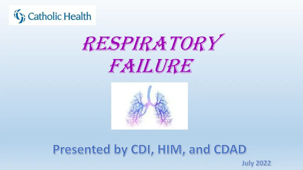
Respiratory Failure Presented by CDI, HIM, and CDAD July 2022
Denials What is a Denial? Every patient has a claim sent to their Insurance Carrier which specifically outlines all care rendered for treatment of Principle diagnosis, secondary diagnoses, and any procedures. The insurance company reviews the claim and all diagnoses submitted, then performs a thorough review of the medical record to ensure all diagnoses are well supported, monitored, and treated accordingly. When insurance company reviews the record and finds lack of clinical support, conflicting documentation, lack of treatment for a diagnosis etc., they notify the hospital organization via a denial letter that they are removing a diagnosis from the claim and state their reasons for doing so.
Appeal Process Denial letter is reviewed by Clinical Denials and Appeals Team, followed by thorough review of the medical record. Along with assistance from the Physician Advisor, an appeal letter is generated stating our case as to why Catholic Health feels the diagnosis is valid and supported in the medical record. The Appeal letter includes specifics taken right from the documentation in the medical record along with all supportive clinical indicators. It is sent to the Insurance Carrier within 30 days of receipt of denial letter. No Appeal Process Denial letter is reviewed by the Clinical Denials and Appeals Team, followed by thorough review of the medial record. With assistance from Physician Advisor, if the reviewed record does not have the proper documentation and clinical support to validate the denied diagnosis, Catholic Health does not send an appeal to the Insurance Carrier. In other words, Catholic Health is in agreement with the Insurance Carrier that the stated diagnosis is not clinically supported.
If Appealed, Appeal letter is sent to Insurance Carrier. If the Insurance Carrier: Disagreeswith Catholic Health Agreeswith Catholic Health Original diagnosis that was denied is re-instated and Catholic Health receives full value for resources associated with patient encounter. Catholic Health receives the appropriate severity of illness, risk of mortality, and more accurate length of stay for patient encounter. More accurate reflection of the Hospital s CMI (Meaning your are caring for very sick patients) Catholic Health can choose to do no further appeal, and the diagnosis is removed from the patient s claim. Catholic Health can choose to try second review with the insurance company. If the second review is still upheld Catholic Health can choose to send for a third review to an Outside company. The Outside Company does a thorough review of the patient s record hearing both sides, and makes a non-biased decision on the validity of the diagnosis. Whomever the decision is in favor of, the other must pay the Outside Company for their services.
When an Appeal : IS LOST Diagnosis is removed from patient s claim. This indicates you never took care of patient for this during their stay. This leads to Catholic Health returning resources to insurance company. This leads to lowering of patient s SOI/ROM for said visit, which also lowers what their length of stay should have been. This lowers the Hospital s CMI (Case Mix Index). A low CMI indicates you are not caring for very sick patients.
We Are Here to Help You! CODING CDAD CDI Review the medical record and all documentation while patient is in the hospital. Review the medical record and all documentation after the patient is discharged. Review the documentation after the Insurance Carrier has denied a diagnosis. Final codes record and sends to billing department for final claim to the Insurance Carrier. Place queries/talk with the Provider to obtain clarification, more specific documentation and to alert Provider of conflicting and/or inaccurate documentation. Team up with the Physician Advisor to effectively determine medical necessity, to locate and identify crucial medical record documentation in order to support the denied diagnosis and Appeal to the Insurance Carrier. Also query if there is inaccurate, conflicting documentation or if further clarification is needed in the medical record. Goal is to obtain a complete and accurate record before the patient is discharged. Goal is to obtain a complete and accurate record.
Patient Encounters where Insurance Carrier Denied Acute Respiratory Failure
Case Scenario 1 Admitted for Acute hypoxic respiratory failure secondary to RLL Pneumonia, likely community acquired; Reactive airways, bronchospasm CC: difficulty breathing EMS: pale, dusky nail beds . SpO2 90% RA placed on 2 L/min NC, SpO2 improved to 95% ED Vitals: 97.2, 84, RR 16- 22; 150/60: SpO2 98% 2L/min; RR 16-20 during inpatient stay Imaging: Consolidative process RLL, infiltrate such as PNA Labs: WBC 14.7.. No blood gas obtained ER RN: Resp Assess: cough, SOB, unlabored; taken off O2 -> SpO2 down to 89%; Placed back on 2 L/min up to 96% ER MD: still having significant hypoxia and on re-exam, wheezed on forced expiration . Physical exam: pulmonary effort normal, No respiratory distress. Admit for hypoxic respiratory failure and PNA Treatments: breathing treatments (SpO2 s remained upper 80s, low 90s even after breathing treatments), IV solumedrol, IV rocephin, IV azithromycin H&P: physical exam: mild tacypneic, mild labored breathing though is currently off NC, O2 92% on RA; No accessory muscle use. RLL mild rhonchi, very faint end expiratory wheeze scattered.. Nursing notes: Respiratory WDL SpO2 s ranging 94-98% on room air Taken off Oxygen at end of ED stay, and remained off oxygen rest of hospitalization.
Is the Diagnosis of Acute hypoxic Respiratory Failure supported? ER MD documented to admit for Acute hypoxic respiratory. His Physical exam: pulmonary effort normal, no respiratory distress, but adds, still having significant hypoxia and on re-exam, wheezes on forced Expiration Answer is NO ER RN: SOB, Unlabored breathing SpO2 90% on room air Why is it not supported: 1. Physical Exam did not reflect a patient in acute respiratory failure 2. Providers did not document their supportive clinical indicators for the acute hypoxic respiratory failure 3. There is no clinical support based on the documentation in the record 4. Patient was not refractory to oxygen SpO2 increased to 98% on 2LPM; Not on O2 at home Was taken off O2 SpO2 down to 89% so placed back on 2 LPM SpO2 back up to 96% Was weaned off O2 in ER, Sats 94-98% room air H&P Physical Exam: No accessory muscle use; Captures acute hypoxic respiratory failure due to the Pneumonia. Daily progress notes have Acute hypoxic respiratory failure documented Discharge Summary has acute hypoxic respiratory failure as a DC Diagnosis Documentation was consistent LOS: 3 days
Case Scenario 2 53 year old male with no PMH, cc: headache, dizziness, intermittent cough (flu like symptoms), shortness of breath, joint/muscle pain, low grade fever, and no appetite. Vitals: Temp: 102.1 (oral); 168/94- 98- 22. SpO2 89% on room air at rest, 88% upon ambulation/exertion. Improved to 94% on 2 lpm O2 via NC Nursing Physical Assessments: shortness of breath at rest and cough that is non-productive . Also documenting patient s SpO2 s and amount of supplemental oxygen consistently throughout the record. Physician Physical Assessments: hypoxia, 89% r/a. Moderate respiratory distress, labored breathing, tachypnea, unable to complete full sentences . Diminished lung sounds, is uncomfortable, ill appearing . Ambulatory pulse ox 88% on room air; ABG pH 7.53 pCO2 28 pO2 176 CT Chest : groundglass densities H&P: Admitted for Sepsis, bilateral Pneumonia, and hypoxia. 89% on r/a, 88-89% with ambulation, increasing to 94 -96% on 2L NC.. Treat pneumonia and continue supplemental oxygen Patient treated with IV Rocephin and Zithromax, IV fluids, tylenol Physicians Documenting in all general medical PN: acute hypoxic respiratory failure requiring continual oxygen supplement and still hypoxic and still SOB, hypoxic on exertion O2 titrated up from 2L to 3L NC and then again to 4 lpm Attempted to titrate off of oxygen but did not tolerate Discharge Summary: Diagnosed and treated for Sepsis secondary to bilateral multifocal pneumonia: Acute hypoxic respiratory failure secondary to bilateral pneumonia. Discharging home on 2 L of O2 via NC . Acute hypoxic respiratory failure diagnosis DENIED by Insurance company Not Supported
Appeal Response from Catholic Health Hospital Course: 53 year old with no past medical history presents to ER form urgent care with complaints of excruciating headache, low grade fever, dizziness, cough, and chills. Patient notes he had flu symptoms last week which initially improved and have now worsened. In ER he was found to be hypoxic with RA sat 89% with mild- moderate respiratory distress and mild tachypnea requiring supplemental oxygen. WBC 14.2 ABG at 1921: pH 7.53 PC02 28 Vital Signs in ER: 1811 168/94 98 20 T102.1 89% on RA 2011 148/94 92 22 T99.3 90% on RA 2108 94% on 02 2lnc (p/f ratio 260) 2052 88% on RA with ambulation 2222 145/95 89 22 96% on 02 2lnc ER MD: 2241: on exam mild respiratory distress noted with labored breathing and mild tachypnea. Unable to complete full sentences. Moderate rales. +wheezing. 2245: hypoxia on arrival with 02 sat 89% on RA. Mild to moderate respiratory distress with inability to complete full sentences. CT of chest consistent with bilateral pneumonia. On arrival to floor at 2040 patient was still hypoxic with 02 sat of 93% on 3lnc which is a p/f ratio of 212.Oxygen Increased to 4 LPM NC. He was noted to have persistent hypoxia requiring supplemental oxygen with multiple p/f ratios <300 throughout hospitalization indicating hypoxic respiratory failure. He was unable to be weaned to room air as patient continued to desaturate to <90% and was discharged with new home oxygen. H&P, Daily progress notes and Discharge Summary: all consistently documenting Acute hypoxic respiratory failure with documentation of persistent hypoxia, requiring increase in oxygen (up to 4L), and will be going home on supplemental oxygen. Documented the cause of the acute hypoxic respiratory failure by linking it to patient s Pneumonia
Outcome Insurance carrier reversed the denial, informing acute hypoxic respiratory failure was valid. Why did we win this denial case? Physical exam captured appropriately by all Providers documentation of labored breathing, tachypnea, respiratory distress, unable to speak in complete sentences. CONSISTENT DOCUMENTATION of Acute hypoxic respiratory failure - captured in H&P, daily progress notes, and Discharge Summary with supportive indicators, treatment and patient s response to treatment. (remains persistently hypoxic despite interventions, O2 needing increase to 4L; will need to go home on supplemental oxygen) Patient was refractory to 2-3 lpm oxygen.. Saturations remained low on 2L, requiring increase in oxygen to 4L, documented P/F ratios
So, When it Comes to the Patients Medical Record Your documentation is looked at by many to make sure it is accurate, valid, and complete. Be cautious when using cut and paste. You do not want to carry forward any inaccurate or old information. It can be seen by all. Make sure your physical exams give a clear picture of what the patient looks like at the time you are completing it. Be sure to individualize and update your documentation daily to ensure the documentation is accurate.
Acute Respiratory Failure A life threatening condition that requires immediate and aggressive management Develops over minutes to hours A syndrome of inadequate gas exchange: oxygenation and/or carbon dioxide elimination The individual displays evidence of some sort of increased work of breathing (ie. Use of accessory muscles, tachypnea (respirations > 20/min), labored respirations, unable to speak in sentences, retractions, cyanosis, tripod position, dyspnea) respiratory distress must be present and symptoms patient is displaying must be documented Patient is refractory to 2-3 lpm supplemental oxygen via nasal cannula Requires intervention with respiratory treatments and either invasive mechanical ventilation, or non- invasive modalities such as NRB, BiPAP/CPAP, Venti mask, higher flow oxygen (4 lpm and higher) Must document/identify the underlying cause of patient s Acute Respiratory Failure
Types of Acute Respiratory Failure Acute respiratory failure with hypoxia lack of oxygen in your arterial blood (PaO2 < 60), CO2 normal/close to normal A. Clinical Findings/Physical Exam: restlessness, tachycardia, irregular heartbeat, tachypnea, dyspnea, profuse sweating, cyanosis of skin, fingertips, or lips; pursed lip breathing, use of accessory muscles, retractions, metabolic acidosis B. Underlying causes: COPD exacerbation, Pneumonia, Pulmonary Edema, CHF, other acute diseases of the lung C. Low pulse oximetry readings (SpO2 <91% room air) & refractory to 2-3 lpm supplemental oxygen via nasal cannula; ABG PaO2 <60 D. Treatments: Duonebs, steroids, HFNC, NRB, Venti-mask, Bipap/Cpap, mechanical ventilation
Types of Acute Respiratory Failure Acute respiratory failure with hypercapnia Failure of lungs to eliminate adequate CO2 resulting in an accumulation of carbon dioxide (CO2) in arterial blood (PaCO2 > 50 mm Hg) and a pH < 7.35 A. Clinical Findings/Physical Exam: anxiousness, daytime sluggishness, a change in mental status, somnolence, short shallow breathing/shortness of breath, tachycardia, headache and respiratory acidosis B. Underlying causes: sedative overdose, COPD, Interstitial lung disease, Stroke, brainstem disease C. Obtain ABG: acute respiratory acidosis (pH < 7.35) & PaCO2 is elevated >50mmHg D. Treatments: Bipap/Cpap; HFNC
Chronic Hypoxic/Hypercapnic Respiratory Failure Condition in which the inability to effectively exchange carbon dioxide and oxygen results in chronic low oxygen levels, and chronic high carbon dioxide levels. Develops slowly over time and progresses as patient s disease progresses. Patient has a severe pulmonary condition requiring continuous use of oxygen (ie.. End stage COPD, Interstitial lung disease) No significant deviation from baseline ABGs when measured Patient s display shortness of breath, especially on exertion, wheezing, coughing up mucous, fatigue, anxiety, daily headache, bluish skin, lips, nailbeds These patient s are on oxygen around the clock to maintain SpO2 of 91%. Often accompanied by compensatory metabolic acidosis (elevated HCO3, normal pH)
Home Use of Supplemental Oxygen PRN use and nocturnal use of oxygen is NOT chronic hypoxic respiratory failure. Patient needs to be oxygen dependent. Oxygen around the clock = oxygen dependence IS chronic hypoxic respiratory failure. If Oxygen dependent, be sure to document a diagnosis of chronic hypoxic respiratory failure.
Acute on Chronic Respiratory Failures Acute on chronic respiratory failure with hypoxia Already on oxygen around the clock due to underlying condition Increased dyspnea, treated with an increase in their normal dose of supplemental oxygen SpO2 < 91% on chronic oxygen administration. Acute on chronic respiratory failure with hypercapnia pCO2 > 50mm Hg with a pH < 7.35, or, Increase in baseline pCO2 by > 10 mm Hg When determining an acute on chronic respiratory failure, one must understand the patient s already compromised baseline function. If the baseline is unknown, it can be retrospectively compared once the acute illness is resolved and added treatments have been discontinued.
Postoperative Respiratory Failure Inability to wean from mechanical ventilation within 48 hours of surgery Is considered a pulmonary complication from the surgical procedure- MD MUST DOCUMENT IT IS A DIRECT COMPLICATION OF THE SURGERY Postoperative Respiratory Failure Unplanned intubation/re- intubation in the postoperative period Must identify underlying cause (ie.. PNA. COPD/Bronchospasm, CHF, Atelectasis, Anesthetics, Opioids, Sedatives, Atelectasis
When to document Postoperative Respiratory Failure You cannot wean patient from Ventilator within 48 hours after surgery OR Unplanned Intubation or Reintubation in the postoperative period AND You are able to link the Respiratory Failure directly to the Surgical Procedure
Patients on Mechanical Ventilation for: 1. Airway Protection Needs Clarification: A. Was patient intubated just as a precaution (to be on the safe side). If so, do not capture a diagnosis of Acute Respiratory Failure as it would not be clinically valid. B. Or, is patient truly in Acute Respiratory failure by displaying respiratory signs and symptoms such as agonal breathing, increased secretions causing concern for Aspiration etc. If so, be sure to document the respiratory dysfunctions that led to the Acute Respiratory Failure diagnosis. 2. Postoperative Rest If patient remains on mechanical ventilation during the postoperative period for rest and sedation, patient is not in Acute Respiratory Failure. Therefore, do not capture a diagnosis of Acute Respiratory Failure as it would not be clinically valid.
ABG Analysis Single most important lab test for evaluation of respiratory failure Determines how effectively the lungs are delivering oxygen to the blood and how efficiently the lungs are eliminating carbon dioxide from the blood Also helps indicate how well the lungs and kidneys are working together to maintain normal blood pH (acid-base balance) Acute hypoxic respiratory failure Chronic hypoxic respiratory failure Acute hypercapnic respiratory failure Chronic hypercapnic respiratory failure pH normal normal decreased Near normal paCO2 Normal- decreased Normal- decreased Increased (>50 mm Hg) Increased (>50 mm Hg) paO2 Decreased (<60 mm Hg) Decreased (<60 mm Hg) Decreased (<60 mm Hg) Decreased (<60 mm Hg) HCO3 normal normal normal increased
P/F Ratio Objective tool to identify and confirm acute hypoxic respiratory failure at any time while patient is receiving supplemental oxygen Used mainly in the Clinical Denials and Appeals Process Indicates what the PaO2 would be on room air if oxygen discontinued Taken from blood gas measurements P represents the PaO2 (arterial pO2) from the ABG F represents the FiO2 percent of inspired O2 that the patient is receiving expressed in a decimal (ie. 40% oxygen = FiO2 of 0.40) P/F Ratio < 300 is equal to a pO2 < 60 mm on room air
VGB Analysis Alternative to ABG when evaluating patient s with metabolic or respiratory disturbances Safer and less painful technique than an ABG Assists in quick determination if your patient is hypercapnic by estimating pCO2 Can be criticized for their lack of accuracy: A VGB pO2 does not correlate well with ABG paO2. You need to estimate the paO2 using the SpO2. Are unreliable in patient s with shock and certain medical conditions.
It is not Acute Respiratory Failure if: The patient comes in with a SpO2 <91% in no respiratory distress, is not on home O2, and needs to be placed on 2-3L supplemental oxygen. As long as maintains good saturations on the 2-3L, and improves with treatment of the acute condition, this is Hypoxemia. The patient shows no signs of any respiratory distress, and is not refractory to 2-3L oxygen via nasal cannula (are breathing normal/comfortably on this dose) Not being on home oxygen and requiring oxygen during inpatient stay is not an automatic acute respiratory failure diagnosis. Patient must meet all criteria (obvious respiratory distress, being refractory to 2-3L oxygen, and needing supplemental oxygen via NRB, Venti-mask, higher flow of O2 via nasal cannula, Bipap/Cpap) for this diagnosis.
When to Document Respiratory Failure: The patient must have a good chance of having progressive breathing dysfunction and failure leading to death if not treated. The patient must demonstrate signs and symptoms of respiratory distress. Very often the History and Physical examinations of hypoxemic patients describe patients, who are breathing easily and are in no distress. Patients with acute respiratory failure don t breathe easily unless they are markedly hypercarbic and on the verge of total respiratory failure. The patient must show hemodynamic signs of respiratory dysfunction. Very often the History and Physical examinations of hypoxemic patients describe patients as having normal vital signs and breath sounds. Patients with acute respiratory failure must show abnormalities of vital signs like tachycardia, tachypnea, and exaggerated paradoxical pulse. They need to show abnormal breath sounds and low airflow such as low peak expiratory flow rates, and abnormal abdominal thoracic muscle movements. The patient must be on significant amounts of supplemental oxygen, higher flow O2 (4 lpm or more), BiPAP, etc. Putting a patient on 2-3 liters of oxygen, and having them then be normal does not suggest acute respiratory failure
When is it only Hypoxemia? Po2 <60 on ABG HYPOXIA OR SpO2 <=91% on room air WHILE Maintaining SpO2 > 91% on 2-3 lpm O2 via nasal cannula WITH NO RESPIRATORY DISTRESS Hypoxemia is not equal to acute respiratory failure. You can have hypoxemia without having acute respiratory failure. A good example is obstructive sleep apnea. Patients can have profound hypoxemia, and yet have no respiratory failure.
Documentation Requirements When documenting Respiratory Failure you must specify/document: Acuity: Is it Acute, Acute on Chronic, or Chronic Acute hypoxic respiratory failure Respiratory Failure Type: Is it hypoxic, hypercapnic, or both hypoxic and hypercapnic Physical Symptoms: ie. Labored breathing, tachypnea, use of accessory muscles, unable to speak in full sentences, retractions, air hunger Pulmonary: respiratory distress, tachypnea, labored respirations, accessory muscle usage Pulmonary: Effort Normal, No respiratory distress
Documentation Requirements When documenting Respiratory Failure you must specify/document: Supportive Clinical indicators: ABG findings, SpO2, use of Venti-mask, NRB, Bipap/Cpap, P/F ratio, hypoxic on supplemental oxygen, refractory to 2-3 L oxygen requiring increase in oxygen The underlying cause of patient s Respiratory Failure How the patient is responding to treatments Update status in the daily progress notes: improving, resolving, worsening, resolved. Include the diagnosis in H&P, daily progress notes, and Discharge summary CONSISTENCY!
Conclusion/Recap! Patient must display physical symptoms and be refractory to oxygen for a diagnosis of acute hypoxic respiratory failure. Make sure physical assessment is in accordance with someone in acute hypoxic respiratory failure. If pulmonary effort normal, and no distress seen, it is unlikely patient is in respiratory failure. If patient has SpO2 < 91% but responds to supplemental oxygen applied without upward titration, patient has hypoxemia, and NOT Acute hypoxic respiratory failure Not being on oxygen at home, and then having O2 applied in the hospital setting due to hypoxia/SOB does not equate to an acute hypoxic respiratory failure diagnosis. Patient must meet criteria. PRN, Nocturnal use of oxygen IS NOT chronic hypoxic respiratory failure.
Conclusion/Recap! Use of oxygen around the clock ISchronic hypoxic respiratory failure. Document the clinical support and underlying cause of patient s acute hypoxic respiratory failure. If patient s are kept on ventilator after a surgical procedure for rest, airway protection, it is not appropriate to document Postoperative Respiratory failure. Only document reason patient is being maintained on the ventilator (ie. Rest), or if in true Acute Respiratory Failure, be sure patient meets all clinical criteria and include the supportive indicators in your documentation. Postoperative Respiratory Failure If unable to wean the patient from ventilator after 48hours, or patient requires intubation/re-intubation in the postoperative period, and MD links the respiratory failure to the procedure in the documentation.
LASTLY There are no ICD-10 codes for: 1. Type 1 Respiratory Failure 2. Type 2 Respiratory Failure 3. Type 3 Respiratory Failure 4. Type 4 Respiratory Failure ** they will go to an unspecified respiratory failure . Therefore, in order to accurately capture patient s severity of illness, be sure to capture these diagnosis in terms of: 1. Acute hypoxic respiratory failure 2. Acute hypercapnic respiratory failure 3. Postoperative Respiratory Failure 4. Acute Respiratory failure secondary to Shock (cardiovascular instability) YOU WILL BE QUERIED
Please feel free to contact us with Questions Clinical Documentation Integrity(CDI) Deborah Mazur Manager, Clinical Documentation Integrity Office #: (716) 601-3752 Email: dmazur@chsbuffalo.org Coding Education Quality Auditing Justine Whitman Manger, Coding Education Quality Auditing Inpatient Office #: (716) 447-6581 Email: Jnolder@chsbuffalo.org Clinical Denials and Appeals Department (CDAD) Brandon Schulz-Koller Director, Documentation Cycle Integrity and Improvement Office #: (716) 601-3602 Email: bkoller@chsbuffalo.org