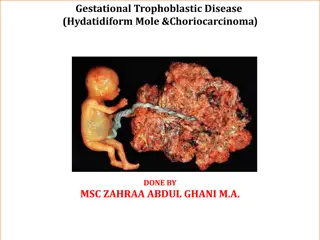
Understanding Gestational Trophoblastic Diseases (GTD) and Hydatidiform Mole
Learn about Gestational Trophoblastic Diseases (GTD) including definitions, classifications, metastatic/non-metastatic diseases, and the incidence and etiology of Hydatidiform Mole. Discover the types and prevalence of GTD as well as risk factors associated with this condition.
Uploaded on | 0 Views
Download Presentation

Please find below an Image/Link to download the presentation.
The content on the website is provided AS IS for your information and personal use only. It may not be sold, licensed, or shared on other websites without obtaining consent from the author. If you encounter any issues during the download, it is possible that the publisher has removed the file from their server.
You are allowed to download the files provided on this website for personal or commercial use, subject to the condition that they are used lawfully. All files are the property of their respective owners.
The content on the website is provided AS IS for your information and personal use only. It may not be sold, licensed, or shared on other websites without obtaining consent from the author.
E N D
Presentation Transcript
GESTATIONAL TROPHOBLASTIC DISEASES (GTD) DR. AMIT KUMAR Associate Professor (Department Of OBG) Madhav Homeopathic Medical College, Abu Road, Sirohi
DEFINITION: Gestational Trophoblastic Disease (GTD) encompasses a spectrum of proliferative abnormalities of trophoblasts pregnancy. Persistent GTD (persistently raised - hCG) is referred as gestational trophoblastic neoplasia (GTN). associated with
CLASSIFICATION OF GTD Hydatidiform mole: Complete Partial Invasive mole Placental site trophoblastic tumor Choriocarcinoma
Non metastatic disease (confined to the uterus) Metastatic disease: A. Low risk (good prognosis) Disease is present < 4 months duration Initial serum hCG level < 40,000 mIU/ml Metastasis limited to lung and vagina No prior chemotherapy No preceding term delivery
B. High risk (poor prognosis) Long duration of disease (> 4 months) Initial serum hCG > 40,000 mIU/ml Brain or liver metastasis Failure of prior chemotherapy Following term pregnancy WHO score > 8
HYDATIDIFORM MOLE (Syn: Vesicular mole) DEFINITION: It is an abnormal condition of the placenta where there are partly degenerative and partly proliferative changes in the young chorionic villi. These result in the formation of clusters of small cysts of varying sizes. TYPES: Complete Incomplete (partial)
Complete Incomplete (partial)
INCIDENCE -The highest incidence is in Philippines being 1 in 80 pregnancies and lowest in European countries 1 in 752 and USA being about 1 in 2000. The incidence, in India, is about 1 in 400. ETIOLOGY: Its prevalence is highest in teenage pregnancies and in those women over 35 years of age. The prevalence appears to vary with race and ethnic origin.
Faulty nutrition caused by inadequate intake of protein, & Low dietary intake of carotene is associated with increased risk. Disturbed maternal immune mechanisms suggested by (a) Rise in gammaglobulin level in absence of hepatic disease (b) Increased association with AB blood group which possesses no ABO antibody.
The higher the ratio of paternal: maternal chromosomes, the greater is the molar change. Complete moles show 2: 0 paternal/maternal ratio whereas partial mole shows 2: 1 ratio. History of prior hydatidiform mole increases the chance of recurrence (1 to 4%).
SYMPTOMS Vaginal bleeding Varying degree of lower abdominal pain may be due to The patient becomes sick without any apparent reason. Vomiting of pregnancy becomes excessive Breathlessness Expulsion of grape like vesicles per vaginam is diagnostic of vesicular mole. History of quickening is absent.
INVESTIGATIONS: Full blood count, ABO and Rh grouping. Hepatic, renal and thyroid function tests are carried out. Sonography: Characteristic pregnancy is snowstorm appearance Definitive diagnosis is made by histological examination of the products of conception features of molar
DIFFERENTIAL DIAGNOSIS: Threatened abortion Fibroid or ovarian tumor with pregnancy Multiple pregnancy:
COMPLICATIONS OF MOLAR PREGNANCY (1) Hemorrhage and shock (2) Sepsis (3) Perforation of the uterus (4) Pre-eclampsia with convulsion on rare occasion (5) Acute pulmonary insufficiency
MANAGEMENT The principles in the management are: Suction evacuation (SE) of the uterus as early as the diagnosis is made. Supportive therapy: Correction of anemia and infection, if there is any. Counseling for regular follow-up
PARTIAL OR INCOMPLETE MOLE In partial hydatidiform mole, the affection of the chorionic villi is focal. There is a fetus or at least an amniotic sac. Microscopic examination of the dilated chorionic villi shows predominant hyperplasia of the syncytiotro-phoblast and presence of fetal blood vessels with fetal red blood cells.
Once the diagnosis is made and the fetus is not alive,termination of pregnancy is to be done. Even if the fetus is alive, the patient should be warned about the risks involved to the fetus if pregnancy is continued.
PLACENTAL SITE TROPHOBLASTIC TUMOR (PSTT) It is a rare histological diagnosis. Syncytotrophoblastic cells are generally absent. So there is persistent low level of serum or urinary hCG. The tumor arises from the intermediate trophoblasts of the placental bed and is composed mainly of cytotrophoblastic cells. Patient presents with vaginal bleeding. Local invasion into the myometrium and lymphatics occurs. PSTT is not responsive to chemotherapy. Hysterectomy is the preferred treatment.


