Explore the process of hemoglobin synthesis, the role of erythropoietin, and factors influencing hemoglobin production. Learn about anemia, its causes, and how vitamins and trace metals play a crucial role in blood health.
Please find below an Image/Link to download the presentation.
The content on the website is provided AS IS for your information and personal use only. It may not be sold, licensed, or shared on other websites without obtaining consent from the author. If you encounter any issues during the download, it is possible that the publisher has removed the file from their server.
You are allowed to download the files provided on this website for personal or commercial use, subject to the condition that they are used lawfully. All files are the property of their respective owners.
The content on the website is provided AS IS for your information and personal use only. It may not be sold, licensed, or shared on other websites without obtaining consent from the author.
Objectives: 1- Quantitative determination of hemoglobin in a blood sample. 2- Importance of glucose 6-phosphate dehydrogenase (G6PD) activity in erythrocytes (hemolysate?). 3- Qualitative determination of hemoglobin S (HbS) in blood using a phosphate solubility method.
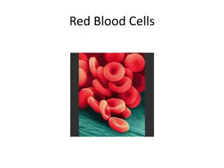
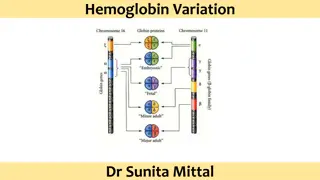
Hemoglobin: Hemoglobin the RBCs (erythrocytes) that transport oxygen from the lungs to the rest of the body and carbon dioxide back to the lungs. (Hb),is the iron-containing oxygen-transport metalloprotein in The protein part: 4 subunits of globin protein. The non-protein part: heme group (iron + Protoporphyrin). Hb is made up of protein and non-protein parts.
Hemoglobin Synthesis The circulation blood of normal adult contain about 750 g of Hb and of this about 7 8 g are degraded daily. The globin part (protein), of Hb can be reutilized only after catabolism into its constituent amino acid. The free heam, is broken down into bile pigment which is excreted. Iron alone is reutilized in the synthesis of Hb. Some factors which can influence the rate of Hb synthesis (Rate of RBC formation): Erythropoietin (EPO) Levels. Iron Availability, If iron levels are low, the rate of Hb synthesis may be reduced. Iron is obtained from the diet and is absorbed in the small intestine. Bone Marrow Health. Nutritional Status. Oxygen Demand, body requires increased oxygen-carrying capacity, such as at high altitudes or during strenuous physical activity, the rate of Hb synthesis may increase to meet the demand. (anoxia, hypoxia?)
Erythropoietin is a glycoprotein hormoneformed in kidney in response to decrease oxygen carrying capacity (hypoxia or anoxia), in order to stimulate the erythropoiesis.
Role of Vitamins, Trace Metals and Cofactors: 1- Biotin, pantothenic acid, pyridoxal phosphate and coenzyme A; are essential coenzymes required for the synthesis of haem. The deficiency of pyridix I phosphate plays a role in human disease while the deficiency of folic acid can cause megaloblastic anemia. 2- Trace metals, e.g. copper and cobalt are known to play a role. Copper is playing a role in the absorption of ion, while Cobalt is an essential constituent of vitamin B12. 3- One common cause of megaloblastic anemia is a deficiency in vitamin B12 or folic acid. Megaloblastic anemia is diagnosed through blood tests that reveal abnormalities in the size and shape of red blood cells, as well as testing the levels of vitamin B12 and folic acid in the blood.
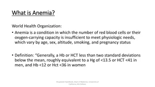
Anemia : Is a medical condition characterized by a decrease in the number of red blood cells or a decrease in the amount of hemoglobin in the blood. Hemoglobin, is a protein in red blood cells that carries oxygen from the lungs to the body's tissues. There are several types and causes of anemia, including: 1- Iron-deficiency anemia. 2- Vitamin deficiency anemia. 3- Hemolytic anemia: this type of anemia, red blood cells are destroyed or removed from the bloodstream earlier than normal, leading to a decreased number of red blood cells. 4- Aplastic anemia. 5- Sickle cell anemia.
Glucose-6-phosphate dehydrogenase, in individuals, with defect in glucose-6- phosphate dehydrogenase, causes red blood cells to break down prematurely. This destruction of red blood cells is called hemolysis. The most common medical problem associated with glucose-6-phosphate dehydrogenase deficiency is hemolytic anemia, which occurs when red blood cells are destroyed faster than the body can replace them. This type of anemia leads to paleness, yellowing of the skin and whites of the eyes (jaundice), dark urine, fatigue, shortness of breath, and a rapid heart rate.
Importance of G6PD G6PD is the enzyme responsible for the initial deviation of glucose into pentose phosphate pathway to form 6-phosphogluconate. This pathway provides NADPH2 in the erythrocyte for the conversion of oxidized to reduced glutathione and for other reactions such as reduction of methemoglobin(ferric (Fe3+) state). Deficiency of G6PD: 1- Hemolytic anemia. 2- Jaundice: Hemolysis of red blood cells can release a pigment called bilirubin, which can accumulate in the body and cause yellowing of the skin and eyes (jaundice). 3- Hemoglobinuria: In severe cases of G6PD deficiency, the breakdown of red blood cells can release hemoglobin into the urine, resulting in a condition called hemoglobinuria. This can cause the urine to appear dark or tea-colored.
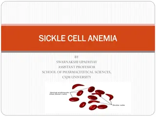
Sickle cell anemia , It is a genetic disorder in which the red blood cells have an abnormal shape, leading to their premature destruction and a reduced oxygen-carrying capacity. Hemoglobin S (Hgb S), is an abnormal type of hemoglobin that you can inherit from parents. It is the type present in the sickle cell anemia. - In sickle cell anemia, the amino acid present at position 6 in the chain of hemoglobin S (HbS) is valine. The substitution of valine for glutamic acid at position 6 is what causes the abnormal structure and function of HbS, leading to the sickling of red blood cells in individuals with sickle cell anemia. - Hgb S, causes red blood cells to become stiff and abnormally shaped. Instead of having a normal round, disk shape, these red blood cells become sickle-shaped, or crescent-shaped. These cells don't live as long as normal red blood cells. Because of their shape, they get stuck inside small blood vessels. These problems cause symptoms of sickle cell disease.
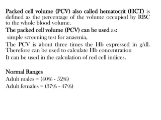
Estimation of blood hemoglobin: Principle: The ferrouse (Iron II), in each heme in RBC is oxidized by ferricyanide to Fe(III)- methemoglobin . A cynide group (CN-) is then attached to the iron atom ((Fe3+) state because it is positively charge), to give the brown cyanmethemoglobin which can be estimated quantitatively by reading the absorbance at 540 nm. Normal Hb conc.: for men: 14 - 18 g/dl, for women : 12 - 16 g\dl Level of Hb is associated with polycythemia and dehydration. Level of Hb is associated with anemia.
Principle: Erythrocytes are lysed (by saponin). The released hemoglobin is reduced (by dithionite) in phosphate buffer. Reduced HbS is characterized by its very low solubility , So that in the presence of HbS, the solution become turbid and the lines behind the test tube will not be visible while, if no HbS was present the clear solution will permit the lines to be seen through the test tubes.


 in blood.")