Pancreatitis is inflammation of the pancreas that can vary in severity from mild to life-threatening. Acute pancreatitis is reversible, while chronic pancreatitis involves irreversible damage. Common causes include alcoholism, gallstones, and metabolic disorders. Learn more about the etiological factors and manifestations of pancreatitis.
Please find below an Image/Link to download the presentation.
The content on the website is provided AS IS for your information and personal use only. It may not be sold, licensed, or shared on other websites without obtaining consent from the author. If you encounter any issues during the download, it is possible that the publisher has removed the file from their server.
You are allowed to download the files provided on this website for personal or commercial use, subject to the condition that they are used lawfully. All files are the property of their respective owners.
The content on the website is provided AS IS for your information and personal use only. It may not be sold, licensed, or shared on other websites without obtaining consent from the author.
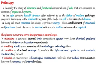
Pancreatitis Inflammation of the pancreas. The clinical manifestations can range in severity from a mild, self-limited disease to a lifethreatening acute inflammatory process The duration of the disease can range from a transient attack to an irreversible loss of function.
Pancreatitis Acute or chronic In acute pancreatitis, gland can return to normal if underlying cause of the pancreatitis is removed. By contrast, chronic pancreatitis is defined by irreversible destruction of exocrine pancreatic parenchyma.
Acute pancreatitis Acute pancreatitis is a group of reversible lesions characterized by inflammation of the pancreas ranging in severity from edema and fat necrosis to parenchymal necrosis with severe hemorrhage. 80% of cases in western countries are associated with one of two conditions: biliary tract disease or alcoholism. Gallstones are present in 35% to 60% of cases of acute pancreatitis.
Acute pancreatitis Obstruction of the pancreatic duct system eg. periampullary tumors, congenital cystic dilatation of the common bile duct, biliary "sludge," and parasites (particularly Ascariasis lumbricoides and Clonorchis sinensis organisms) Medication More than 85 drugs have been reported to cause acute pancreatitis. e.g . thiazide diuretics, estrogens, etc Metabolic disorders Including hypertriglyceridemia, hyperparathyroidism, and other hypercalcemic states Acute ischemia Vascular thrombosis, embolism, vasculitis and shock Trauma: Blunt trauma Iatrogenic injury during surgery or endoscopic retrograde cholangiopancreatography
Acute pancreatitis: Morphology The morphology of acute pancreatitis ranges from inflammation and edema to severe extensive necrosis and hemorrhage.
Acute pancreatitis: Morphology The basic alterations are (1) microvascular leakage causing edema (2) necrosis of fat by lipolytic enzymes (3) an acute inflammatory reaction (4) proteolytic destruction of pancreatic parenchyma (5) destruction of blood vessels with subsequent interstitial hemorrhage Fat necrosis results from enzymatic destruction of fat cells. The released fatty acids combine with calcium to form insoluble salts that precipitate .
Acute pancreatitis Pathogenesis. autodigestion of the pancreatic substance by inappropriately activated pancreatic enzymes. Thus, activation of trypsinogen is an important triggering event in acute pancreatitis.
Acute pancreatitis: Clinical Features. Abdominal pain is the cardinal manifestation of acute pancreatitis. Full-blown acute pancreatitis is a medical emergency . These patients usually have the sudden onset of an "acute abdomen . Characteristically, the pain is constant and intense and is often referred to the upper back. There is leukocytosis, hemolysis, disseminated intravascular coagulation, fluid sequestration, acute respiratory distress syndrome, and diffuse fat necrosis. Peripheral vascular collapse and shock with acute renal tubular necrosis may occur
Acute pancreatitis Laboratory findings: marked elevation of serum amylase levels during the first 24 hours, followed within 72 to 96 hours by a rising serum lipase level.
Acute pancreatitis The key to the management is "resting" the pancreas by total restriction of food and fluids and by supportive therapy. Most patients recover fully. About 5% die from shock during the first week of illness. Acute respiratory distress syndrome and acute renal failure are fatal complications. In surviving patients, sequelae include a sterile pancreatic abscess and a pancreatic pseudocyst.
Chronic pancreatitis Chronic pancreatitis is characterized by inflammation of the pancreas with destruction of exocrine parenchyma, fibrosis, and, in the late stages, the destruction of endocrine parenchyma. The chief distinction between acute and chronic pancreatitis is the irreversible impairment in pancreatic function that is characteristic of chronic pancreatitis.
Chronic pancreatitis There is significant overlap in the causes of acute and chronic pancreatitis. By far the most common cause of chronic pancreatitis is long-term alcohol abuse and biliary tract disease, and these patients are usually middle-aged males.
Chronic pancreatitis Less common causes of chronic pancreatitis include the following: Hypercalcemia, hyperlipidemia. Long-standing obstruction of the pancreatic duct by pseudocysts, calculi, trauma, neoplasms, or pancreas divisum. Tropical pancreatitis, which is a poorly characterized disease seen in Africa and Asia. It has been attributed to malnutrition. Hereditary pancreatitis Idiopathic chronic pancreatitis.
Chronic pancreatitis: Morphology Chronic pancreatitis is characterized by parenchymal fibrosis, reduced number and size of acini with relative sparing of the islets of Langerhans, and variable dilation of the pancreatic ducts These changes are usually accompanied by a chronic inflammatory infiltrate around lobules and ducts. Grossly: gland is hard, sometimes with extremely dilated ducts and visible calcification
Chronic pancreatitis: Clinical Features Silent or repeated attacks of abdominal pain or persistent abdominal and back pain. Attacks may be precipitated by alcohol abuse, overeating , or the use of opiates and other drugs. During an attack of abdominal pain, there may be mild fever and mild-to-moderate elevations of serum amylase. Calcifications can be seen within the pancreas by CT scan and ultrasonography.
Chronic pancreatitis: Clinical Features Complications: Severe pancreatic exocrine insufficiency Chronic malabsorption Diabetes mellitus (due to destruction of islets of Langerhans) Severe chronic pain Pancreatic pseudocysts
PSEUDOCYSTS OF PANCREAS Pseudocysts are localized collections of necrotic- hemorrhagic material rich in pancreatic enzymes. Such cysts lack an epithelial lining (hence the prefix "pseudo"), and they account for majority of cysts in the pancreas. Pseudocysts usually arise after an episode of acute pancreatitis, or of chronic alcoholic pancreatitis.
PSEUDOCYSTS OF PANCREAS Morphology. Pseudocysts are usually solitary. Pseudocysts can range in size from 2 to 30 cm in diameter. While many pseudocysts spontaneously resolve, they may become secondarily infected, and larger pseudocysts may compress or even perforate into adjacent structures. They can produce abdominal pain and predispose to intraperitoneal hemorrhage or peritonitis.