Upper GI Bleeding and Portal Hypertension
Upper gastrointestinal bleeding (UGIB) and portal hypertension are critical medical conditions associated with significant mortality rates. Causes, risk factors, prognosis, history, physical examination, and work-up of UGIB are discussed in detail, highlighting the importance of early evaluation, proper triage, and management strategies.

Download Presentation

Please find below an Image/Link to download the presentation.
The content on the website is provided AS IS for your information and personal use only. It may not be sold, licensed, or shared on other websites without obtaining consent from the author.If you encounter any issues during the download, it is possible that the publisher has removed the file from their server.
You are allowed to download the files provided on this website for personal or commercial use, subject to the condition that they are used lawfully. All files are the property of their respective owners.
The content on the website is provided AS IS for your information and personal use only. It may not be sold, licensed, or shared on other websites without obtaining consent from the author.
E N D
Presentation Transcript
Upper GI Bleeding&Portal Hypertension Dr. Ali Jaffer Upper GI surgeon
Background Bleeding derived from a source proximal to the ligament of Treitz. Bleeding from the upper GI tract is approximately 4 times more common than bleeding from the lower GI tract. Mortality rates from UGIB are 6-10% overall. Comorbid diseases increase the death rate. Rebleeding and continued bleeding is a significant factor of mortality.
Aetiology Causes of upper gastrointestinal bleeding. Condition Ulcers Oesophageal Gastric Duodenal Erosions Oesophageal Gastric Duodenal Mallory Weiss tear Oesophageal varices Tumour Vascular lesions, e.g. Dieulafoy s disease 0.5 Others % 60 6 21 33 26 13 9 4 4 4 0.5 5
Prognosis The following risk factors are associated with an increased mortality, recurrent bleeding, the need for endoscopic hemostasis, or surgery: 1. Age older than 60 years 2. Severe comorbidity 3. Active bleeding (eg, witnessed hematemesis, red blood per nasogastric tube, fresh blood per rectum) 4. Hypotension 5. Red blood cell transfusion greater than or equal to 6 units 6. Inpatient at time of bleed 7. Severe coagulopathy Patients who present in hemorrhagic shock have a mortality rate of up to 30%
History Important information potential comorbid conditions, medication history, and potential toxic exposures severity, timing, duration, and volume of the bleeding Hematemesis Melena Hematochezia Syncope Dyspepsia Epigastric pain Heartburn Diffuse abdominal pain Dysphagia Weight loss Jaundice
Physical Examination The goal; to evaluate for shock and blood loss. Assessing the patient for hemodynamic instability and clinical signs of poor perfusion is important early in the initial evaluation to properly triage patients with massive hemorrhage. Worrisome clinical signs and symptoms of hemodynamic compromise include Tachycardia of more than 100 beats per minute (bpm) Systolic blood pressure of less than 90 mm Hg, Cool extremities, syncope, and other obvious signs of shock, Ongoing brisk hematemesis, The occurrence of maroon or bright-red stools, which requires rapid blood transfusion. Pulse and blood pressure should be checked with the patient in supine and upright positions to note the effect of blood loss. Significant changes in vital signs with postural changes indicate an acute blood loss of approximately 20% or more of the blood volume. Signs of chronic liver disease should be noted, including spider angiomata, gynecomastia, splenomegaly, ascites, ....etc. nodular liver, an abdominal mass, and enlarged and firm lymph nodes.
Work up Assessment of hemorrhagic shock patients who present in hemorrhagic shock have a mortality rate of up to 30% Estimated Fluid and Blood Losses in Shock Class 1 Class 2 Class 3 Class 4 Blood Loss, mL Up to 750 750-1500 1500-2000 >2000 Blood Loss,% blood volume Up to 15% 15-30% 30-40% >40% Pulse Rate, bpm < 100 >100 >120 >140 Blood Pressure Normal Normal Decreased Decreased Normal or Increased Respiratory Rate Decreased Decreased Decreased Urine Output, mL/h >35 30-40 20-30 14-20 CNS/Mental Status Slightly anxious Mildly anxious Anxious, confused Confused, lethargic
Work up Hemoglobin Value and Type and Crossmatch Blood CBC should be checked frequently (4-6h) during the first day. The patient should be crossmatched for 2-6 units, based on the rate of active bleeding. Patients with significant comorbid conditions (eg, advanced cardiovascular disease) should receive blood transfusions to maintain myocardial oxygen delivery to avoid myocardial ischemia. The more units required, the higher the mortality rate. Operative intervention is indicated once the blood transfusion number reaches more than 5 units. Coagulation Profile The patient's prothrombin time (PT), activated partial thromboplastin time (PTT), and international normalized ratio (INR) should be checked to document the presence of coagulopathy. The coagulopathy may be consumptive and associated with a thrombocytopenia
Work up Endoscopy Diagnostic&theraputic Endoscopy should be performed immediately after endotracheal intubation (if indicated), hemodynamic stabilization, and adequate monitoring in an intensive care unit (ICU) setting have been achieved. Chest Radiography Computed Tomography Scanning Liver disease for cirrhosis, pancreatitis with pseudocyst and hemorrhage, aortoenteric fistula, and other unusual causes of upper GI hemorrhage. Nuclear Medicine Scanning Nuclear medicine scans may be useful in determining the area of active hemorrhage. Angiography Angiography may be useful if bleeding persists and endoscopy fails to identify a bleeding site. Transcatheter arterial embolization (TAE) should be considered for all patients with a known source of arterial UGIB that does not respond to endoscopic management, with active bleeding and a negative endoscopy. Nasogastric Lavage PPIs Treatment of underlying cause
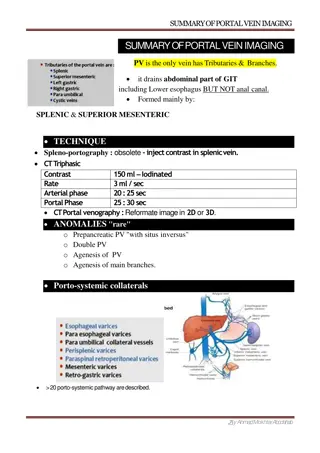
Portal Hypertension The portal venous system contributes approximately 75% of the blood and 72% of the oxygen supplied to the liver. In the average adult, 1000 to 1500 mL/min of portal venous blood is supplied to the liver. The normal portal venous pressure is 5 to 10 mmHg, and at this pressure, very little blood is shunted from the portal venous system into the systemic circulation. As portal venous pressure increases, the collateral communications with the systemic circulation dilate, and a large amount of blood may be shunted around the liver and into the systemic circulation. Lower oesophagus; Left gastric veins (portal system) -> lower branches of oesophageal veins (systemic veins) Upper part of anal canal; Superior rectal veins (portal) -> inferior and middle rectal veins (systemic) Umbilicus; Paraumbilical veins (portal) -> epigastric veins (systemic) Area of the liver; Intraparenchymal branches of right division of portal vein (portal) -> retroperitoneal veins (systemic) Hepatic and splenic flexures; Omental and colonic veins (portal) -> retroperitoneal veins (systemic)
Aetiology of portal hypertension Sinusoidal Intrahepatic Cirrhosis Viral infection Alcohol abuse Primary biliary cirrhosis Autoimmune hepatitis Primary sclerosing cholangitis Metabolic abnormality Postsinusoidal Intrahepatic Vascular occlusive disease Posthepatic Budd-Chiari syndrome Congestive heart failure Inferior vena caval web Constrictive pericarditis Presinusoidal Sinistral/extrahepatic Splenic vein thrombosis Splenomegaly Splenic arteriovenous fistula Intrahepatic Schistosomiasis Congenital hepatic fibrosis Nodular regenerative hyperplasia Idiopathic portal fibrosis Myeloproliferative disorder Sarcoid Graft-versus-host disease Portal hypertension per se produces no symptoms, it is usually diagnosed following presentation with decompensated chronic liver disease and encephalopathy, ascites or variceal bleeding.
Management of bleeding varices General resuscitation Medical emergency ICU Two large pore peripheral canulae Resuscitation, avoid fluid overload (why?) Correction of coagulopathy; Vit K(10mg) i.v., tranexamic acid (1g i.v), FFP, platelet transfusion Activation of major blood transfusion protocol Drug therapy (terlipressin) splanchnic vasocinstriction Prophylactic antibiotics Endoscopy ; 50% PHT non variceal bleeding Sengstaken-Blakemore, temporary control; Once inserted, the gastric balloon is inflated with 300 mL of air and retracted to the gastric fundus, where the varices at the oesophagogastric junction are tamponaded by the subsequent inflation of the oesophageal balloon to a pressure of 40 mmHg. The balloons should be temporarily deflated after 12 hours to prevent pressure necrosis of the oesophagus.
Management of bleeding varices Endoscopic treatment of varices Endoscopic band ligation Endoscopic sclerotherapy Transjugular intrahepatic portosystemic stent shunts the main treatment of variceal haemorrhage that has not responded to drug treatment and endoscopic therapy. Complications: Liver capsule perfuration . Intraperitoneal hemorrhage Occlusion resulting in further variceal bleeding Post shunt encephalopathy 40% of cases TIPS stenosis (50% after one year) Surgical shunts for variceal haemorrhage Surgical shunts are an effective method of preventing rebleeding from oesophageal or gastric varices, as they reduce the pressure in the portal circulation by diverting the blood into the low- pressure systemic circulation. Long-term -blocker therapy and chronic sclerotherapy or banding are the main alternatives. Liver transplantation is the only therapy that will treat both portal hypertension and the underlying liver disease.
Ascites Portal vein thrombosis is a common predisposing factor to the development of ascites in chronic liver disease. In patients without evidence of liver disease, malignancy is a common cause Aspiration of the peritoneal fluid allows the measurement of protein content to determine whether the fluid is an exudate or transudate, an amylase estimation to exclude pancreatic ascites. Cytology will determine the presence of malignant cells Microscopy and culture will exclude primary bacterial and tuberculous peritonitis. Treatment of ascites in chronic liver disease Salt restriction Diuretics Abdominal paracentesis TIPSS Liver transplantation