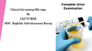
Urinalysis
Urinalysis is a crucial diagnostic tool used to assess various conditions affecting the urinary system. Understanding the physical and chemical properties of urine helps in diagnosing diseases like diabetes, renal failure, and urinary tract infections. This practical guide covers the interpretation of urinalysis results, the importance of dipsticks, and correlations with clinical findings. Explore the normal and abnormal parameters of urine, including color, odor, volume, pH, protein, glucose, ketones, nitrite, and bilirubin levels. Enhance your knowledge of urinalysis for accurate disease identification and patient management.

Download Presentation

Please find below an Image/Link to download the presentation.
The content on the website is provided AS IS for your information and personal use only. It may not be sold, licensed, or shared on other websites without obtaining consent from the author.If you encounter any issues during the download, it is possible that the publisher has removed the file from their server.
You are allowed to download the files provided on this website for personal or commercial use, subject to the condition that they are used lawfully. All files are the property of their respective owners.
The content on the website is provided AS IS for your information and personal use only. It may not be sold, licensed, or shared on other websites without obtaining consent from the author.
E N D
Presentation Transcript
King Saud University College of Medicine Department of Pathology Clinical Biochemistry unit Urinalysis
By end of the this practical, you should be able to: Understand the physical and chemical examinations of urine in health and disease. Perform urinalysis using Dipsticks. Recognize the value of urinalysis as a tool for diagnosis of diseases e.g. metabolic diseases, kidney disorders and for urinary tract infections (UTIs). Interpret the results of urinalysis and correlate it with the patient s clinical findings.
Urine: Urine is a fluid excreted by most of mammals including humans. It is formed in the kidneys (renal glomeruli). The fluid undergoes chemical changes before it is excreted as urine. Normal urine excretion by a healthy person is about 1.5 L per day.
Physical Properties of urine PARAMETER NORMAL ABNORMAL POSSIBLE CAUSES Diabetes, chronic renal failure Dehydration, Acute renal failure Presence of pus cells, bacteria, salt or epithelial cells Polyuria Oligouria Volume 0.4-2.0 L/day Appearance Clear Cloudy Excessive fluid intake, uncontrolled DM, chronic renal failure Dehydration, carotenoid ingestion Jaundice Blood, drugs etc Methemoglobin, alkaptonuria, melanoma, black water fever glomerulonephritis Diabetic ketoacidosis Contaminated and long standing exposed urine Phenylketonuria Maple syrup urine disease Colorless Orange Yellow-Green Red Colour Pale Yellow Dark brown-black smoky Fruity Ammoniacal Mousy Burnt sugar Crystals, salts or cells Odor Urineferous Blood clots, necrotic tissues and urinary stones Deposits None ketosis (diabetes mellitus & starvation), severe diarrhea, metabolic and respiratory acidosis, excessive ingestion of meat and certain fruits Acidic Reaction (pH) 4.6 - 7.0 Respiratory and metabolic alkalosis, Urinary tract infection, Vegetarians Alkaline
Chemical Properties of urine PARAMETER NORMAL ABNORMAL POSSIBLE CAUSES Nephrotic syndrome, glomerulonephritis,, multiple myeloma, lower UTI, tumors or stones Protein < 200mg/day Proteinuria Uncontrolled DM, gestational diabetes, Fanconi s syndrome Glucose None Glucosuria Diabetic ketoacidosis, Glycogen storage disease, starvation, Prolonged vomiting, Unbalanced diet: high fat & Low CHO diet Ketones None Ketonuria UTI Nitrite None Detected Bilirubin None Detected Hepatic and post-hepatic jaundice Normal Trace (1mg/dl) Urobilinogen > 2 mg/dl Jaundice Acute & chronic glomerulonephritis, Trauma , cystitis , renal calculi and tumors, Bleeding disorders (Hemophilia). Hematuria Blood None Hemoglobinopathies, Malaria, Transfusion reaction (Blood Incompatibility) Hemoglobinuria
Proteins Normally less than 200 mg protein is excreted in the urine daily; more than this level leads to a condition called Proteinuria . Glomerular proteinuria: It is due to glomerular permeability filtration of high molecular weight proteins ( e.g. glomerulonephritis). Tubular proteinuria: It occurs as a result of tubular reabsorption with normal glomerular permeability excretion of low molecular weight proteins (e.g. chronic nephritis)
Nephrotic syndrome Large amounts of protein are lost in the urine and hypoproteinaemia develops. Increase protein excretion in urine can be one of the following two types: A: High molecular weight protein excretion: Glomerular proteinuria due to increase glomerular permeability leading to filtration of high molecular weight proteins B: Low molecular weight protein excretion: Tubular proteinuria due to decrease reabsorption with normal glomerular permeability
Urinalysis (using dipstick): Principle: Dipsticks are plastic strips impregnated with chemical reagents which react with specific substances in the urine to produce color-coded visual results. They provide quick determination of pH, protein, glucose, ketones, urobilinogen, bilirubin, blood, hemoglobin, nitrite, and specific gravity. The depth of color produced relates to the concentration of the substance in urine. Color controls are provided against which the actual color produced by the urine sample can be compared .The reaction times of the impregnated chemicals are standardized.
Procedure: Dip the strip in the urine sample provided then remove it immediately. Remove the excess urine and keep the strip in a horizontal position. Read the color produced within 30-60 seconds (Color changes after more than 2 minutes are of no significance). Match the color changes to the color scale provided. Give a full report about: - Physical examination - Chemical examination
Case I (Urine Sample I) A 12-year-old girl, a known patient with T1DM, presented to Emergency drowsy with short history of vomiting and abdominal pain. On examination: - Tachycardia - Tachypnea with a fruity smell of breath. - BP: 85/50 mmHg (Ref range: 100/66-135/85 mmHg) - Blood sugar: 26.7 mmol/L (Ref range: 3.9-5.6 mmol/L) - HbA1C: 9.9% (Ref range: 5.7-6.4%) - Blood pH: 7.1 (Ref range: 7.35 7.45) - Circulating Ketone bodies: Positive A mid stream Urine sample was collected for complete urinalysis. 1- Do urinalysis using dipsticks and give a full report regarding: A- Physical examination. B- Chemical examination. 2- What is the most likely diagnosis?
Case II (Urine Sample II) A 49-old woman with a history of DM came to hospital with fever, weakness and dysuria (pain during urination) for the last three days. The results of her laboratory tests were as follows: Test Result Reference range 3.9-5.8 mmol/L 55-120 mmol/L 2.5-6.4 mmol/L 135-145 mmol/L 3.5-5.1 mmol/L Fasting blood glucose Creatinine Urea Sodium Potassium 7.5 75 3.7 140 3.9 A mid stream Urine sample was collected for complete urinalysis. Microscopic examination of urine showed:- WBCs: > 100/HPF (Ref range: 2-3/HPF ) RBCs: 50/ HPF (Ref range: 0-2/HPF ) 1- Do urinalysis using dipsticks and give a full report regarding: A- Physical examination. B- Chemical examination. 2- What is the most likely diagnosis?
Case III (Urine Sample III) A 6-year-old boy, developed marked edema over a period of few days. His mother had noticed puffiness around the eyes, characteristically in the morning. She also noticed that his urine had become frothy. His general practitioner ordered the following investigations: Test Result 58 3.4 136 4.0 34 14 11 15 Reference range 55-120 mmol/L 2.5-6.4 mmol/L 135-145 mmol/L 3.5-5.1 mmol/L 60-80 g/L 35-50 g/L 3.2-5.2 mmol/L 0.5-2.27 mmol/L Creatinine Urea Sodium Potassium Total protein Albumin Cholesterol Triglycerides A mid stream Urine sample was collected for complete urinalysis. 1- Do urinalysis using dipsticks and give a full report regarding: A- Physical examination. B- Chemical examination. 2- What is the most likely diagnosis?
Task I. Physical Examination : Appearance Color . Odor . Deposits . Specific gravity . Reaction ( pH)
Task II. Chemical Examination: Item Observation Comment Protein Glucose Ketones Nitrite Bilirubin Urobilinogen Blood


:")

")
")
")