Viral Infections of Respiratory Tract Lecture 3 Summary
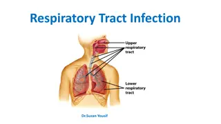
In this lecture, the focus is on viral infections of the respiratory tract, covering commonly encountered viruses like Influenza, Rhinovirus, Coronavirus, Para-influenza viruses, Respiratory Syncytial viruses, and Adenovirus. Details on Orthomyxoviruses, Influenza A, B, and C are provided, including their pathogenesis, clinical syndromes, immunity, and laboratory diagnosis methods. Learn about the different types of influenza viruses, complications, and symptoms associated with respiratory infections.

Download Presentation

Please find below an Image/Link to download the presentation.
The content on the website is provided AS IS for your information and personal use only. It may not be sold, licensed, or shared on other websites without obtaining consent from the author.If you encounter any issues during the download, it is possible that the publisher has removed the file from their server.
You are allowed to download the files provided on this website for personal or commercial use, subject to the condition that they are used lawfully. All files are the property of their respective owners.
The content on the website is provided AS IS for your information and personal use only. It may not be sold, licensed, or shared on other websites without obtaining consent from the author.
E N D
Presentation Transcript
VIRAL INFECTIONS OF THE RESPIRATORY TRACT Lucture 3
The commonest of human infection 1-Influenza virus >Orthomyxoviridae 2-Rhinovirus>Picornaviridae family 3-Coronavirus>Coronaviridae family 4-Para influenza viruses> Paramyxoviridae family 5-Respiratory Synctial viruses >Paramyxoviridae 6-Adenovirus> Adenoviridae family.
1-Orthomyxoviruses Influenza Virus 1)Single, Stranded negative sense RNA with 8 helical segments, This virus is highly susceptible to mutations and rearrangements within the infected host. 2)Helical capsid symmetry 3)Enveloped viruses which contains 2 projecting glycoprotein spikes. Heamagglutinin HA The virus can agglutinate certain erythrocyte. Neuroamindase NA from infected cell. attachment. an enzyme help in releasing progeny virus formation
*Very Important Types of Influenza Viruses Influenza A Hard to control it Influenza B Influenza C Infect human Infect human only Infect human and Cause outbreaks&epidemic Cause mild illness animals. Can cause epidemic Antigenicdrift only pandemic and pandemic epizootic. epidemic Antigenic drift Out break antigenic shift.
Pathogenesis & Immunity: local upper respiratory tract infection. According to the immunity, it can cause localized infection or spread to the lower respiratory tract infection. Viremia usually& occurs (fever) . self limiting condition in Immunocompetent person. Complication of Influenza: Primary Influenza Pneumonia. 2ndbacterial-pneuomonia > Strep. pneumoniae, H.influenzae Myositis (inflammation of the muscle). Post influenza encephalitis.(brain) Bronchial Asthma. Sinusitis.
Clinical Syndrome: Transmission inhalation of respiratory secretion Incubation period 1 - 4 days Seasonal variation usually in winter self limiting disease prognosis Symptoms: Sudden onset of fever Malaise Headache Sneezing sore throat It takes 3 days. Non-productive cough
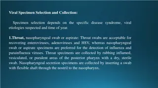
Laboratory Diagnosis: Clinical diagnosis. Laboratory investigation done to distinguish influenza viruses from other respiratory viruses and to identify the type and strain. Specimen: Nasopharyngeal aspirate, nasal washing Culture: on primary Monkey Kidney cytopathic effect occur 2- 3 days. Rapid and direct detection of influenza virus A or B from nasopharyngeal aspirate by immunofluorescence and ELISA(most common laboratory diagnosis) Rapid antigen immunofluorescence assay :Assay performed on cells from a nasopharyngeal aspirate, showing typical nuclear and cytoplasmic apple-green fluorescence after staining with monoclonal antibodies specific for influenza A. RT-PCR (Nucleic acid testing)
Treatment: Amantadine: Is only effective against influenza A virus(inhibiting the un coating step of influenza A virus.) It has both therapeutic and prophylactic .It significantly reduced the duration of fever and illness is given to high risk group of patients who are not vaccinated because they have allergy from egg.
Oseltamivir (Tamiflu) : It is Neuraminidase inhibitor which help the influenza virus invade respiratory tract cells. It has to be given within the first 48 hours after the exposure. INFLUANZA VACCINE Tow types of vaccine ,both contain the current influenza A & B . Vaccine should be given in October or November ,before the influenza season begins. Yearly booster dose recommended. 1-The Flu shot vaccine Inactivated (Killed vaccine), Given to people older than 6 months, including healthy people as well as high risk groups. 2-The Nasal spray flue vaccine (Flu mist) This is a live attenuated vaccine,Approved for use in healthy people only between 5- 49 years age.
2-RHINOVIRUSES. Common cold accounts for 1/3 to of all acute respiratory infections in humans. Rhinoviruses are responsible for 60% of common colds cases, Common cold is a self-limited illness. More than 100 serologic types of rhinoviruses, No vaccine available. Transmitted directly by respiratory droplet. RHINOVIRUSES (PICORNAVIRUS family) small non enveloped virus(20-30 nm),SS-RNA virus. RHINOVIRUS are acid labile(sensitive).
3-Coronaviruses ssRNA enveloped with positive polarity. second cause of common cold . Symptoms runny nose, sneezing and nasal obstruction, mild sore throat, headache and malaise that last for one week. NO FEVER Clinical presentation of common cold: Laboratory Diagnosis: Usually no need. Complication: Usually due to secondary bacterial infection Treatment and Prevention: 1. Acute sinusitis 2) Acute otitis media. No specific treatment. 3) Exacerbation of chronic bronchitis ,bronchial asthma. No vaccine available. infect several human cells , including lower but not upper respiratory, kidney ,intestinal, and liver cells. Severe Acute Respiratory Syndrome SARS SARS is a viral infection, causes Atypical pneumonia, can infect all age groups, and can lead to death especially among people with existing chronic condition. causative agent of SARS A new mutation of coronavirus.Coronavirus is difficult to isolate and not easily grown in tissue culture. Coronavirus is able to survive in dry air for up to 3 hours, but can be killed by exposure to ultra-violet light.
4-Para Influenza Viruses paramyxoviridae family Enveloped SS RNA,. There are four para influenza viruses: 1, 2, 3, 4 (occur mainly in winter.) Transmitted by respiratory droplets. Envelop surface projection presents as Heamagglutinin HA , Neuroamindase NA, F-glucoprotins which cause cell TO cell membrane to fuse syncytia Clinical Syndromes: 1-Croup or Acute Laryngotracheobronchitis: a56rhom parainfulenza Type I,II seen in infants & young children < 5 years. Harsh cough,difficult inspiration which can lead to airway obstruction which need hospitalization to do tracheotomy. 2-Bronchiolitis and pneumonia: Sometime parainfluenza type 3 can cause bronchiolitis and pneumonia in young children. 3-Common Cold: Seen in older children and adult. 4-Immunocompromized: Parainfluenza type 3 very dangerous, especially in bone marrow transplant patient.
Laboratory Diagnosis: nasopharyngeal aspirate by direct immunofluorescent. Isolation of the virus from nasopharyngeal aspirate OR mouth wash in cell culture will appear as multinucleated giant cell (syncitia). Treatment and Prevention: Hospital admission for infant having Croup for careful monitoring of upper airway (endotracheal intubation and tracheotomy), No specific antiviral treatment, no vaccine available.
5-Respiratory Syncytial Virus (RSV) paramyxoviridae family. Enveloped ,ss RNA . The virus transmitted by respiratory droplets, RSV virus is very contagious with( I.P. 3-6 days) infection mainly in winter. The importance of RSV lies in its tendency to invade the lower respiratory tract of infant <6 months causing >Bronchiolitis& pneumonia. Clinical Syndromes: RSV can cause any respiratory tract illness from commoncold>(0-6m)pneumonia In old children and adult can cause common cold . Bronchiolitis an important and life threatening disease in infant especially under 6 months of life, started with fever, nasal discharge, rapid breathing, respiratory distress and cyanosis, it may be fatal in premature infant or infant with underlying disease or immunocompromised infant, also can lead to chronic lung disease in later life. Pneumonia: also an important and life threatening disease.
Laboratory Diagnosis: Isolation of the virus from nasopharyngeal aspirate OR mouth wash in cell culture will appear as multinucleated giant cell (syncitia). ELISA and immunofluorescent for direct detection from nasopharyngeal aspirate. Treatment and Prevention: Infant (oxygen inhalation). Ribavirin givenby inhalation to treat severe Bronchiolitis and pneumonia. Passive immunization with anti-RSV immunoglobulin is available for premature infant. Hospital staff have to follow control measure No vaccine is available.
6-Family Adenoviridae (Adenoviruses) dsDNA, non-enveloped viruses with 47serogroup, grouped into 6 group from A F. infect epithelial cells lining respiratory tract,conjunctiva, gastrointestinal tract, and genital tract. Viremia may occur after this local replication of the viruses so virus can spread to other visceral organs e.g. Urinary bladder have the tendency to become latent in lymphoid tissue, can be reactivated if immunity is low. Spread and Transmission: Fecal oral - Respiratory via respiratory droplets. -Contaminated instruments at eye clinics. - has been cultured from semen, so can be spread by sexual transmission. Clinical Syndrome: infect children and less commonly infect adult. Reactivation occur if the patient become immunocompromised in children or adult.
The main clinical syndromes: 1. 2. 3. Pharyngo-conjunctival fever: It occurs more often in children and presents with pharyngitis& conjunctivitis and fever 4. Keratoconjunctivitis: (Infection of Cornea and Conjunctiva) It is due to irritation of the eye by a foreign bodies, dust or debris, or contaminated instruments at eye clinic. 5. Acute respiratory tract disease: Fever, cough, pharyngitis and cervical adenitis it is mainly occur in Military recruits serotype 4,7). 6. Pneumonia: Particularly type 3-7 are a significant cause of pneumonia in preschool children which can be followed by residual lung damage. 7. Viral gastro-entrites : diarrhea mainly in young children and infant (serotypes 40 and 41). 8. Mesenteric adenitis and intussusceptions : mainly in children. 9. Acute hemorrhagic cystitis, dysuria and heamaturia. 10. Cervicitis and urethritis ? Sexually Transmitted. 11. Systemic infection in immunocompromised patient. 12. In these group of patient infection become severe as pneumonia or hepatitis it can be primary exogenous infection or reactivation. Acute Febrile pharyngitis: Occur in preschool children , fever nasal congestion and cough (URTI) . Conjunctivitis: Follicular conjunctivitis, can occur as sporadic cases or as an outbreaks .
Laboratory Diagnosis: Specimens: nasopharyngeal aspirate ( respiratory cells), Conjunctival swab and Stool. Mainly the diagnosis by direct detection of viral antigen by Immunofluorescence and ELISA. Treatment, Prevention and Control No specific treatment available Live Oral vaccine used to prevent acute respiratory tract infection for Military recruits [adenovirus serotype 4 7].
MCQs 1.Which one of the following is considered as first cause of common cold? a. Rhinovirus b. Coronavirus c. Adenovirus 2.Which one of the following is structurally non-enveloped? a. Parainfluenza b. Coronavirus c. Rhinovirus 3.Which one of the following is the only virus with dsDNA? a. Adenovirus b. Influenza c. Respiratory Syncitial 4.Avian flu do not usually infect humans. a. T b. F 4.A 3.A 2.C 1.A
Good Luck Done by : Microbiology Team contact us: microbiology434@gmail.com


 :")


")

")