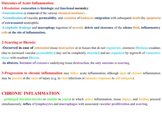
Acute Inflammation Pathogenesis in Images
In this set of images, the pathogenesis of acute inflammation is visually depicted, showcasing processes like exudation, vasodilation, fibrin mesh formation, and inflammation accompanied by necrosis. These detailed images provide a clear insight into the mechanisms underlying acute inflammatory responses.

Download Presentation

Please find below an Image/Link to download the presentation.
The content on the website is provided AS IS for your information and personal use only. It may not be sold, licensed, or shared on other websites without obtaining consent from the author.If you encounter any issues during the download, it is possible that the publisher has removed the file from their server.
You are allowed to download the files provided on this website for personal or commercial use, subject to the condition that they are used lawfully. All files are the property of their respective owners.
The content on the website is provided AS IS for your information and personal use only. It may not be sold, licensed, or shared on other websites without obtaining consent from the author.
E N D
Presentation Transcript
PRACTICAL 2 Inflammation I - Acute Inflammation Foundation Block Pathology Dept, KSU
Pathogenesis of Exudation The process of exudation, aided by endothelial cell contraction and vasodilation, which typically is most pronounced in venules. Collection of fluid in a space is a transudate. If this fluid is protein-rich and has many cells then it becomes an exudate. Foundation Block Pathology Dept, KSU
Exudation in the Alveolar Space Here is vasodilation with exudation that has led to an outpouring of fluid with fibrin into the alveolar spaces along with PMN's indicative of an acute bronchopneumonia of the lung, Foundation Block Pathology Dept, KSU
Exudation of Fibrin in Acute Inflammation Here is an example of the fibrin mesh in fluid with PMN's that has formed in the area of acute inflammation. It is this fluid collection that produces the "tumor" or swelling aspect of acute inflammation. Pathology Dept, KSU Foundation Block
Inflammation with Necrosis - LPF The vasculitis shown here demonstrates the destruction that can accompany the acute inflammatory process and the interplay with the coagulation mechanism. The arterial wall is undergoing necrosis, and there is thrombus formation in the lumen. Foundation Block Pathology Dept, KSU
Inflammation with Necrosis - HPF At higher magnification, vasculitis with arterial wall necrosis is seen. Note the fragmented remains of neutrophilic nuclei (karyorrhexis). Acute inflammation is a non-selective process that can lead to tissue destruction Foundation Block Pathology Dept, KSU
1- Fibrinous Pericarditis Foundation Block Pathology Dept, KSU
Acute Fibrinous Pericarditis - Gross Here, the pericardial cavity has been opened to reveal a fibrinous pericarditis with strands of stringy pale fibrin between visceral and parietal pericardium Pathology Dept, KSU Foundation Block
Acute Fibrinous Pericarditis - Gross Serous fluid at the bottom of the pericardial cavity (arrow) is visible. The epicardial surface appears roughened, compared to its normal glistening appearance; due to the strands of pink-tan fibrin that have formed Foundation Block Pathology Dept, KSU
Acute Fibrinous Pericarditis - Microscopically Lef t Righ t The fibrinous exudate is seen to consist of pink strands of fibrin gutting from the pericardial surface at the upper right .The exudate on the surface is shown enlarged in the inset. Note a considerable number of erythrocytes trapped in the mesh of fibrin threads. Foundation Block Pathology Dept, KSU
Acute Fibrinous Pericarditis - LPF The pericardium is distorted by thick irregular layer of pinkish fibrinous exudate with some red cells and inflammatory cells Foundation Block Pathology Dept, KSU
Acute Fibrinous Pericarditis - HPF The subpericardial layer is thickened by edema and shows dilated blood vessels, chronic inflammatory cells and areas of calcification. Pathology Dept, KSU Foundation Block
2- Acute Appendicitis Foundation Block Pathology Dept, KSU
Normal Appendix - Gross This is the normal appearance of the appendix against the background of the caecum. Foundation Block Pathology Dept, KSU
Acute Appendicitis - Gross Seen here is acute appendicitis with yellow to tan exudate and hyperemia, including the periappendiceal fat superiorly, rather than a smooth, glistening pale tan serosal surface Foundation Block Pathology Dept, KSU
Acute Appendicitis Longitudinal section A case of acute appendicitis : The organ is enlarged and sausage-like (botuliform). This longitudinal section shows the angry red inflamed mucosa with its irregular luminal surface. This appendix does not show late complications, like transmural necrosis, perforation, and abscess formation Foundation Block Pathology Dept, KSU
Acute Appendicitis LPF of the cut section Foundation Block Pathology Dept, KSU
Acute Appendicitis LPF File:Acute Appendicitis, HE 1.jpg Luminal Debris Scattered Neutrophils in the epithelium Lymph Follicle Smooth Muscle layer Foundation Block Pathology Dept, KSU
Acute Appendicitis HPF File:Acute Appendicitis, HE 2.jpg Scattered Neutrophils in the crypt epithelium Foundation Block Pathology Dept, KSU
Acute Appendicitis Histopathology http://www.uaz.edu.mx/histo/pathology/ed/ch_13/c13_s49.jpg This slide shows the muscle layer of the appendix which is permeated with numerous polymorphonuclear leukocytes Foundation Block Pathology Dept, KSU
3- Acute Cholecystitis Foundation Block Pathology Dept, KSU
Acute Cholecystitis Gross Mucocele, stone obstructed the neck , distended , aspiration done and removed by lap chole Foundation Block Pathology Dept, KSU
Acute Cholecystitis Histopathology HPF The neutrophils are seen infiltrating the mucosa and submucosa of the gallbladder in this patient with acute cholecystitis and right upper quadrant abdominal pain with tenderness on palpation Foundation Block Pathology Dept, KSU
4- Skin Pilonidal Sinus Foundation Block Pathology Dept, KSU
Foreign Body Reaction (Pilonidal Sinus) A pilonidal sinus is a sinus tract which commonly contains hairs. It occurs under the skin between the buttocks (the natal cleft) a short distance above the anus. Usually runs vertical between the buttocks and rarely occurring outside the coccygeal region. Foundation Block Pathology Dept, KSU
Foreign Body Reaction (Pilonidal Sinus) Surgically excised pilonidal sinus tracts Foundation Block Pathology Dept, KSU
Pilonidal Sinus Histopathology LPF http://blog.yimg.com/2/DBjAjeN7s59ri3UwzNZdPsXsnsNm.WVZBovrrtkHi6cxsrnpEkewlw--/52/l/YyZhJEo5rTjKgIzDVrF5nQ.jpg The lumen of the sinus and wall contain large number of hair shafts with foreign body giant cells, lymphocytes , macrophages & neutrophils Pathology Dept, KSU Foundation Block
II - Chronic Inflammation Foundation Block Pathology Dept, KSU
1- Chronic cholecystitis with stones Foundation Block Pathology Dept, KSU
Chronic cholecystitis with Gall Stones Gross appearance of gallbladder after sectioning longitudinally. Notice thickness of gall bladder wall, abundant polyhedric stones and small papillary tumor in the cystic duct. Foundation Block Pathology Dept, KSU
Chronic cholecystitis - Histopathology Irregular mucosal folds and foci of ulceration in mucosa. Wall is penetrated by mucosal glands which are present in muscle coat ( Rokitansky- Aschoff sinuses). All layers show chronic inflammatory cells infiltration and fibrosis. Pathology Dept, KSU Foundation Block
Chronic cholecystitis - Histopathology The mucosa is atrophic, with a single layer of flattened epithelium. There is proteinaceous fluid adherent to the mucosal surface, with some bile stained orange-brown crystals toward the upper left in the lumen. The lamina propria shows fibrosis and contains a mononuclear cell infiltrate (small dark blue nuclei). The muscle is hypertrophied compared to normal gallbladder. Foundation Block Pathology Dept, KSU
2- Brain abscess Foundation Block Pathology Dept, KSU
Brain Abscess - gross Cavity contains neutrophils, necrotic cells, bacteria and fibrinous material Central cavity CT of a cerebral abscess. There is a liquefactive center with yellow pus surrounded by a thin wall. Abscesses usually result from hematogenous spread of bacterial infection, but may also occur from direct penetrating trauma or extension from adjacent infection in sinuses Foundation Block Pathology Dept, KSU
Brain Abscess - microscopy Center of the abscess: Cavity contains neutrophils, necrotic cells, bacteria and fibrinous material This trichrome stain demonstrates the light blue connective tissue in the wall of an organizing cerebral abscess. Normal brain is at the right and the center of the abscess at the left. Foundation Block Pathology Dept, KSU
3 - Granulation tissue Foundation Block Pathology Dept, KSU
Granulation Tissue - LPF Section of fragments of edematous, loose connective tissue shows many small newly formed capillaries lined by plump endothelial cells. Proliferation of fibroblasts is seen Foundation Block Pathology Dept, KSU
Granulation Tissue - HPF Inflammatory cells including macrophages, lymphocytes, plasma cells and neutrophils in the oedematous stroma. Pink homogenous collagen fibers may be identified. Foundation Block Pathology Dept, KSU


")
")