Learn about the various methods of fetal assessment used by Dr. Saleh AlAsiri, including early and late pregnancy assessments, rational fetal oxygenation challenges, and monitoring fetal heart activity. These assessments help identify fetal risk factors and ensure optimal care during pregnancy to prevent complications.
Please find below an Image/Link to download the presentation.
The content on the website is provided AS IS for your information and personal use only. It may not be sold, licensed, or shared on other websites without obtaining consent from the author. If you encounter any issues during the download, it is possible that the publisher has removed the file from their server.
You are allowed to download the files provided on this website for personal or commercial use, subject to the condition that they are used lawfully. All files are the property of their respective owners.
The content on the website is provided AS IS for your information and personal use only. It may not be sold, licensed, or shared on other websites without obtaining consent from the author.
E N D
Presentation Transcript
FETAL ASSESSMENT FETAL ASSESSMENT Dr Dr. . Saleh AlAsiri, Saleh AlAsiri, MD, Assistant Professor, Assistant Professor, Consultant Reproductive Endocrinology & Reproductive Endocrinology & infertility Department of Obstetrics & Department of Obstetrics & Gynecology MD, FRCSC, FRCSC, FACOG, FACOG, FACS, Consultant infertility Gynecology FACS, FICS FICS
FETAL ASSESSMENT FETAL ASSESSMENT to identify fetuses at risk of neurologic injury or death in order to prevent it It can be divided into: A-E Early arly pregnancy fetal assessment B-L Late ate pregnancy fetal assessment OR OR A A- - Assessment of low risk pregnancy low risk pregnancy B B - -Assessment of high risk pregnancy high risk pregnancy
RATIONAL fetal oxygenation challenged: - blood flow directed to brain, heart & adrenal & blood flow away from the kidney fetal urine production AF volume. - CNS hypoxia Fetal movement -chemoreceptor's vegally-mediated reflex Fetal heart rate abnormality late deceleration. decrease
EARLY PREGNANCY ASSESSMENT Fetal movement Fetal movement Fetal movement are usually first perceptible to mother ~17w-20w (quickening) 50% of isolated limb movements are perceived 80% of trunk and limb movements Fetal growth Fetal growth Symphysis fundal height (SFH) USS
2- LATE PREGNANCY ASSESSMENT Fetal movement counting (kick chart) Contraction stress test (CST) Non stress test (NST) Doppler Velocimetry (UAV) amniotic fluid index (AFI)
FETAL MOVEMENT COUNTING FETAL MOVEMENT COUNTING It should be started ~ It should be started ~28 &~ &~24 24w in high risk pregnancy w in high risk pregnancy It can reduce avoidable stillbirth It can reduce avoidable stillbirth CARDIFF TECHNIQUE CARDIFF TECHNIQUE - -10 10 movement in movement in 12 - -If abnormal If abnormal further assessment further assessment SADOVSKY TECHNIQUE SADOVSKY TECHNIQUE - -4 4 movement /hour if not felt another hour movement /hour if not felt another hour If not If not further assessment further assessment 28w in normal pregnancy w in normal pregnancy 12 hours hours
CONTRACTION STRESS TEST (CST) Causing uterine contraction over 20 At least 2 2 uterine contractions uterine contractions Uterine contraction restrict O2 delivery to the fetus Normal Normal fetus will tolerate contraction Hypoxic Hypoxic fetus will have late deceleration 20 minutes minutes
NON STRESS TEST (NST) Main advantage over CST is no need for contraction False +ve & false ve higher than CST
NON STRESS TEST NON STRESS TEST The base line 120 Different criteria in fetuses <32w A A- - Reactive Reactive: At least 2 2 accelerations from base line of 15 for at least 15 15 sec sec within 20 B B- - Non reactive Non reactive: No acceleration after 20 minutes another 20 minutes 120- -160 160 beats/minute 15 bpm bpm 20 minutes minutes
NON STRESS TEST ( NON STRESS TEST (NST) NST) If non- reactive in 40 stress test or Biophysical Profile (BPP) 40 minutes contraction
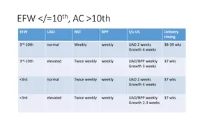
AMNIOTIC FLUID VOLUME ~AFI Amniotic Fluid Index (AFI) -the sum of the maximum vertical fluid pocket diameter in four quarters -the normal value 5 5- -25 25cm cm -<5 : oligohydraminous oligohydraminous ->25 : polyhydraminous polyhydraminous
BIOPHYSICAL PROFILE (BPP) Combines NST , AFV, fetal breathing, body movement & tone. it is a scoring system ( out of 10 it is done over 30 30 min It measure acute hypoxia (NST, body mov. &breathing) & chronic hypoxia (AFI) 10 points)
FETAL BIOPHYSICAL PROFILE/NST+ Normal (score=2) Biophysical Variable 1 episode FBM of at least 30 s duration in 30 min movements Abnormal (score= 0) Absent FBM or no episode >30 s in 30 min 2 or fewer body/limb movements in 30 min Fetal breathing 3 discrete body/limb movements in 30 min Fetal movements 1 episode of active extension with return to flexion of fetal limb(s) or trunk. Opening and closing of the hand considered normal tone Either slow extension with return to partial flexion or movement of limb in full extension Absent fetal movement Either no AF pockets or a pocket<2 cm in 2 perpendicular planes Fetal tone 1 pocket of AF that measures at least 2 cm in 2 perpendicular planes Amniotic fluid volume
BPP The risk of fetal death within 1 week if BPP is normal~ 1/1300
DOPPLER VELOCIMETRY Measurement of blood flow velocities in maternal & fetal vessels Reflect feto-placental circulation Doppler indices from Uterine Artery, Uterine A & Middle cerebral artery In IUGR absent or reversed EDF (end diastolic flow) associated with fetal hypoxia
CHORIONIC VILLUS SAMPLING (CVS): after 10 10w It is the procedure of choice for first trimester prenatal diagnosis of genetic disorders Complication Complication: fetal loss (0.7 CVS procedure and CVS procedure and 1.3 1.3 percent within percent within 30 induced limb defects Second trimester amniocentesis Second trimester amniocentesis is associated with the lowest risk of pregnancy loss; chorionic villus samplings safer than early (ie, before 15 weeks) amniocentesis. Usually done after w first trimester 0.7 percent within percent within 14 30 days) 14 days of a TA days of a TA days), , Procedure- .


")
")


")
")


:")