Fetal Circulation in Embryology: Key Concepts
Fetal circulation involves the unique system that supports blood flow in the fetus. The placenta serves as a crucial respiratory and nutrient exchange organ. Key shunts like Ductus Venosus, Ductus Arteriosus, and Foramen Ovale play vital roles in fetal circulation. The umbilical cord, with its arteries, veins, and unique structures, facilitates the exchange of oxygen and nutrients. Understanding these mechanisms is essential for comprehending fetal development.

Download Presentation

Please find below an Image/Link to download the presentation.
The content on the website is provided AS IS for your information and personal use only. It may not be sold, licensed, or shared on other websites without obtaining consent from the author.If you encounter any issues during the download, it is possible that the publisher has removed the file from their server.
You are allowed to download the files provided on this website for personal or commercial use, subject to the condition that they are used lawfully. All files are the property of their respective owners.
The content on the website is provided AS IS for your information and personal use only. It may not be sold, licensed, or shared on other websites without obtaining consent from the author.
E N D
Presentation Transcript
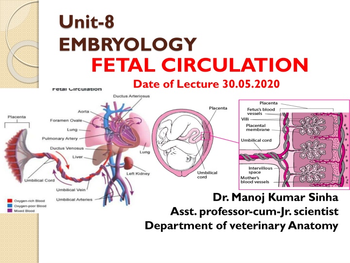
Unit-8 EMBRYOLOGY FETAL CIRCULATION Date of Lecture 30.05.2020 Dr. Manoj Kumar Sinha Asst. professor-cum-Jr. scientist Department of veterinary Anatomy
FETALCIRCULATION The deoxygenated blood and nutritive materials in the fetus is termed as Fetal Circulation The circulatory system of the mother is not directly connected to that of the fetus thus the placenta functions as the respiratory center for the fetus as well as a site of filtration of nutrients and wastages water glucose, amino acids, vitamins etc. freely diffuse through the placenta along with oxygen The uterine arteries carry oxygenated blood to the placenta. circulation of oxygenated blood,
Three shunts are present in fetal life which play important role in fetal circulation Ductus Venosus: connects the umbilical vein to the inferior venacava Ductus Arteriosus: connects the main pulmonary artery to the Aorta Foramen Ovale: Opening between the right and left atrium
Continue.. The placenta accepts deoxygenated blood from the through vessels that leave the fetus through the Umbilical (Umbilical Arteries ) fetus blood Cord The nutrient rich blood then returns to the fetus Umbilical (Umbilical Vein) oxygenated via the cord
UMBILICALCORD Thick cord like structure which extends from center of the abdominal wall of the fetus to the center of the fetal surface of placenta (The structure connecting the fetus with the placenta) umbilical develops from and contains remnants of the yolk sac and allantois The cord
Continue Umbilical cord is made up of : Two umbilical arteries one umbilical vein duct of yolk sac and distal part of allantoic diverticulum Thus umbilical vein carries blood towards the fetus s heart while the umbilical arteries carry blood away All these structures are surrounded by Wharton s jelly( a gelatinous substance made from mucopolysaccharides which protects the blood vessels inside) and finally enveloped by amniotic membrane The Wharton's jelly is formed by the mucoid degeneration of the mesodermal cells of the connecting stalk
Continue Early part of embryonic life two umbilical veins are present , but later only the left vein persists The umbilical vein carrying placental (oxygenated) blood, enters the abdomen of the fetus at the umbilicus and then enters into the liver Left vein then joins the portal vein, hepatic vein and finally opens into the abdominal venacava through Ductus Venosus Then blood conveyed to the right atrium Through Foramen Ovalae blood passes into the left atrium and then to the left ventricle The foramen ovale closes at the time of birth Through the aorta blood goes to the head, fore limbs and small amount of blood passes into the abdominal aorta
Continue From cranial end blood returns through anterior venacava to the right atrium This blood along with blood from abdominal venacava passes into the right ventricle The right ventricular blood goes via pulmonary trunk going to the descending aorta through Ductus Arteriosus because fetal lungs become inactive Ductus Arteriosus is a arterial duct which connects the pulmonary artery with the aorta ( In the fetus there is special connection between the pulmonary artery and the aorta, called Ductus Arteriosus) At birth it becomes occluded but in adult it persists as a ligament (Ligamentum Arteriosum) Some of the blood moves from the aorta through the internal iliac arteries and re-enters the placenta, the maternal circulation
AT THE TIME OF BIRTH Closure of Foramen Ovale Closure of Ductus Arteriosus Closure of Ductus Venosus AFTER BIRTH After birth of the baby the lung, renal, digestive and liver functions are working the fetal circulation undergoes some changes since they are no longer needed