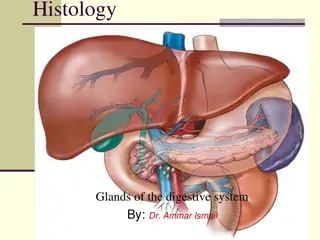
Histology and Biochemistry of the Adrenal Gland Overview
Explore the histology and biochemistry of the adrenal gland, including the stroma, parenchyma divided into cortex and medulla, and the functions of different zones within the cortex. Learn about glucocorticoid investigations, testing methods, and regulatory mechanisms for cortisol secretion. Dive into the intricate details of adrenal gland structure and function.

Uploaded on | 0 Views
Download Presentation

Please find below an Image/Link to download the presentation.
The content on the website is provided AS IS for your information and personal use only. It may not be sold, licensed, or shared on other websites without obtaining consent from the author. If you encounter any issues during the download, it is possible that the publisher has removed the file from their server.
You are allowed to download the files provided on this website for personal or commercial use, subject to the condition that they are used lawfully. All files are the property of their respective owners.
The content on the website is provided AS IS for your information and personal use only. It may not be sold, licensed, or shared on other websites without obtaining consent from the author.
E N D
Presentation Transcript
Histology and biochemistry of the adrenal gland By: abdulaziz abdullah almanie
Adrenal gland It is formed of: 1-Stroma. 2-Parenchyma: that is divided into I. Cortex that is composed of: A-Zona glomerulosa. B-Zona fasciculata. C-Zona reticularis. II. Medulla: It is the central portion of the adrenal gland.It is completely invested with adrenal cortex (not separated from it by CT. septa) It contains: Chromaffin cells (Pheochromocytes): (They produce epinephrine and norepinephrine). 2. Sympathetic ganglion cells :
1- zona glomerulosa:clusters of small columnar cells that are rich in SER and mitochondria. Produces mineralocorticoids e.g. aldosterone hormone 2. zona fasciculata:It is the intermediate and the largest layer of the cortex. *It is formed of columns of large polyhedral cells that are separated by longitudinal sinusoidal capillaries. Its cells secrete glucocoticoids 3. zona reticularis: It is the innermost layer of adrenal cortex.It is formed of anastomosing cords of deep acidophilic cells . The cells secrete androgens.
Glucocorticoid Investigations: 1-Screening tests: assess the clinical diagnosis Include:1-low DXM 2-24-hour urinary free cortisol 2-Confirmatory tests: rule out pseudo- Cushing's syndrome 1-Insulin-induced hypoglycemia: INCREASE CRH Basal serum cortisol: at least 145 nmol/L At 60 - 90 minutes: the level > 425 nmol/L *function Normal values: Serum cortisol: Morning - 7-28 g/dL Afternoon - 2-18 g/dL Plasma ACTH: Morning- 10 - 60 pg/mL Afternoon-5-10 pg/mL *Regulation of ACTH and Cortisol Secretion: 1-Negative feedback control 2-Stress 3-The diurnal rhythm of plasma cortisol Testsused to determine the cause of Cushing's syndrome: 1-Plasma ACTH (Diurnal rhythm) 2-High-dose dexamethasone suppression test *Cortisol and ACTH measurements: 1-Serum [cortisol] and plasma [ACTH] 2-Urinary cortisol excretion (UFC) is < 250 nmol/24 h 3-CRH stimulation test 4-Radiological tests: MRI of pituitary and ultrasound or CT of adrenals