Initial Aflibercept vs. Laser vs. Observation in Diabetic Macular Edema
This study explores the impact of initial treatment with aflibercept, laser, or observation on low-contrast visual acuity in eyes with diabetic macular edema and good vision. It compares different management strategies to evaluate deficits in low-contrast VA and their effects on visual outcomes. The research discusses the importance of contrast sensitivity in driving, recognizing faces, and navigating low-light environments. Findings indicate potential benefits of anti-VEGF therapy in improving contrast sensitivity and vision loss among patients with DME. The study highlights the significance of early intervention and personalized treatment approaches for optimal visual outcomes in diabetic retinopathy.

Download Presentation

Please find below an Image/Link to download the presentation.
The content on the website is provided AS IS for your information and personal use only. It may not be sold, licensed, or shared on other websites without obtaining consent from the author.If you encounter any issues during the download, it is possible that the publisher has removed the file from their server.
You are allowed to download the files provided on this website for personal or commercial use, subject to the condition that they are used lawfully. All files are the property of their respective owners.
The content on the website is provided AS IS for your information and personal use only. It may not be sold, licensed, or shared on other websites without obtaining consent from the author.
E N D
Presentation Transcript
DRCR Retina Network Effect of Initial Aflibercept, Laser, or Observation on Low-Contrast Visual Acuity in Eyes with Diabetic Macular Edema and Good Vision: Ancillary Study Within a Randomized Clinical Trial (Protocol V) 1
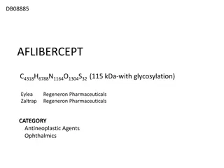
Disclosures Supported by the National Eye Institute and the National Institute of Diabetes and Digestive and Kidney Diseases under award numbers UG1EY014231 and UG1EY023207. Regeneron provided aflibercept for the study and funds to the DRCR Retina Network to defray the study s clinical site costs. 2
Low-Contrast Visual Acuity: Protocol V Low-contrast VA (contrast sensitivity) is important for driving at night, recognizing faces, identifying objects, and seeing in low-light situations DRCR Retina Network Protocol V was a randomized clinical trial of 702 participants (eyes) comparing initial treatment with aflibercept, focal/grid laser, and observation for diabetic macular edema in eyes with high-contrast VA 20/25 or better* Eyes starting with laser or observation could receive aflibercept if VA loss met pre-specified criteria No significant difference between groups in the percentage of eyes with 5-letter high-contrast VA loss from baseline at 2 years 52 of 91 sites comprising 387 of 702 participants (55%) measured LCVA 3 *DRCR Retina Network. Effect of initial management with aflibercept vs laser photocoagulation vs observation on vision loss among patients with diabetic macular edema involving the center of the macula and good visual acuity: A randomized clinical trial. JAMA. 2019;321(19):1880-1894.
Contrast Sensitivity, DME, + Anti-VEGF A small RCT (N = 41 eyes) treated eyes with DME and VA impairment (20/40 to 20/200) with bevacizumab or sham at baseline and 6 weeks* Compared with sham, bevacizumab showed significant improvement in contrast sensitivity at 2 weeks and a non- significant trend at 12 weeks Post hoc analysis of 468 eyes from RIDE/RISE trials showed a mean 2.5-letter improvement in contrast sensitivity at 2 years among eyes treated with ranibizumab** Data from the sham group were not reported * Motta AAL, Bonanomi M, Ferraz DA, et al. Short-term effects of intravitreal bevacizumab in contrast sensitivity of patients with diabetic macular edema and optimizing glycemic control. Diabetes Res Clin Pract. 2019;149:170-178. ** Ip MS, Zhang J, Ehrlich JS. The clinical importance of changes in diabetic retinopathy severity score. Ophthalmology. 2017;124(5):596-603. 4
Objectives 1. Evaluate deficits in low-contrast VA among eyes with good high-contrast VA despite center-involved DME 2. Compare treatment strategies in Protocol V with respect to change in low-contrast VA at 2 years 3. Evaluate correlations of low-contrast VA with high- contrast VA and OCT central subfield thickness 5
Participant Baseline Characteristics Eyes with Baseline Low-Contrast VA Initial Aflibercept N = 112 Initial Laser N = 146 Observation N = 129 62 37% / 63% 91% 7.5 60 Age (y), median Female/Male sex Type 2 Diabetes HbA1c (%), median Race/Ethnicity White Black/African American Hispanic or Latino Other 59 45% / 55% 92% 7.6 32% / 68% 91% 7.7 67% 13% 19% 2% 68% 11% 18% 3% 75% 10% 12% 2% 6
Low-Contrast Visual Acuity Chart ETDRS high-contrast acuity 2.5% low-contrast acuity Waldman AT, Lavery AM, Liu GW, et al. High- and Low-Contrast Letter Acuity Perception Matures With Age in Normally Sighted Children. J Neuroophthalmol. 2020;40(2):148 156. 7
Ocular Baseline Characteristics Eyes with Baseline Low-Contrast VA Initiation With Laser N = 146 Aflibercept N = 112 Observation N = 129 High-Contrast VA Letter Score Mean, letters (Snellen) Low-Contrast VA Letter Score Mean, letters (Snellen) 2 SD below age- specific norms, %* OCT Central Subfield Thickness (Stratus Equivalent) Mean, m 85 (20/20) 85 (20/20) 85 (20/20) 48 (20/125) 45 (20/125) 49 (20/125) 17% 30% 23% 312 317 316 *Pineles SL, Velez FG, Yu F, Demer JL, Birch E. Normative reference ranges for binocular summation as a function of age for low contrast letter charts. Strabismus. 2014;22(4):167-175. 8
Mean Change in Low-Contrast VA Letter Score from Baseline 15 Mean Change in LCVA Letter Score Aflibercept Laser Observation 10 5 +2.7 0 -2.0 -3.1 -5 2 Years: Adjusted Treatment Group Comparisons* Aflibercept vs. Laser:5.3 (-0.2 to 10.8), P = .06 Aflibercept vs. Observation: 5.5 (-0.2 to 11.2), P = .06 Laser vs. Observation:0.2 (-4.6 to 5.0), P = .94 N = 112 N = 146 N = 129 N = 93 N = 117 N = 104 -10 -15 Baseline 1-Year 2-Year Visit *Adjusted for baseline visual acuity and DME treatment in the non-study eye within 4 months before or after randomization. 9 Error bars represent 95% confidence intervals
Impaired Low-Contrast VA % of eyes 2 SD below age-specific norm 35 30 25 20 15 10 5 0 Aflibercept Laser 2 Years Observation Baseline 10 *Pineles SL, Velez FG, Yu F, Demer JL, Birch E. Normative reference ranges for binocular summation as a function of age for low contrast letter charts. Strabismus. 2014;22(4):167-175.
Interpreting Correlations Evans (1996)* proposed the following: .01-.19 very weak .20-.39 weak .40-.59 moderate .60-.79 strong .80-1.0 very strong *Evans, J.D. 1996. Straightforward Statistics for the Behavioral Sciences. Brooks/Cole Publishing; Pacific Grove, Calif.: 1996. 11
High-Contrast VA vs. Low-Contrast VA 2 Years Change from Baseline to 2 Years 100 75 r = 0.47 r = 0.58 Low-Contrast VA Change (95% CI 0.38 to 0.55) Moderate correlation (95% CI, 0.51 to 0.65) Moderate correlation 80 Low-Contrast VA 60 -25 25 40 20 0 -75 50 60 70 80 90 100 High-Contrast VA Change High-Contrast VA 12
Central Subfield Thickness vs. Low-Contrast VA 2 Years Change from Baseline to 2 Years 75 100 r = -0.07 r = -0.20 (95% CI, -0.30 to -0.09) Weak correlation (95% CI, -0.18 to 0.04) No significant correlation Low-Contrast VA Change 80 Low-Contrast VA 60 -300 300 40 20 -75 0 OCT Central Subfield Thickness Change 100 200 300 400 500 OCT Central Subfield Thickness 13
Summary Objective #1: Evaluate deficits LCVA among eyes with good high-contrast VA despite center-involved DME At baseline, among eyes with good HCVA ( 20/25), 23% had a deficit in LCVA After treatment with aflibercept, laser, or observation, only the aflibercept group saw a numeric decline in the percentage of eyes with a deficit in LCVA at 2 years compared with baseline 14
Summary Objective #2: Compare treatment strategies in Protocol V with respect to change in low-contrast VA at 2 years Change in low-contrast VA from baseline was not statistically different between the three treatment strategies Aflibercept vs. Laser 95% CI: -0.2 to 10.8 Aflibercept vs. Observation 95% CI: -0.2 to 11.2 The study was conducted at only a subset of sites in Protocol V and not powered for this objective 15
Summary Objective #3: Evaluate correlations of low-contrast VA with high-contrast VA and OCT central subfield thickness While low-contrast VA and high-contrast VA were moderately correlated, some patients had good HCVA but poor LCVA, which can interfere with overall visual function There was no significant correlation between low-contrast VA and OCT central subfield thickness in cross section at 2 years; there was a weak correlation between changes from baseline 16
Conclusion There was no significant difference between treatment groups in low-contrast VA change from baseline to 2 years Considering the range of the 95% confidence intervals, however, the study may have been underpowered to detect a clinically meaningful benefit between treatment groups 17
Thank You on Behalf of the DRCR Retina Network 18