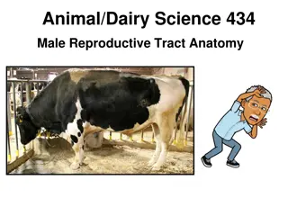
Male Reproductive System - Anatomy and Functions
The male reproductive system consists of primary and secondary sex organs responsible for production and transportation of spermatozoa and male sex hormones. It includes structures like testes, scrotum, reproductive ducts, accessory sex glands, prepuce, and penis. Understanding the anatomy and functions of the male reproductive system is crucial for overall health and fertility.

Download Presentation

Please find below an Image/Link to download the presentation.
The content on the website is provided AS IS for your information and personal use only. It may not be sold, licensed, or shared on other websites without obtaining consent from the author. If you encounter any issues during the download, it is possible that the publisher has removed the file from their server.
You are allowed to download the files provided on this website for personal or commercial use, subject to the condition that they are used lawfully. All files are the property of their respective owners.
The content on the website is provided AS IS for your information and personal use only. It may not be sold, licensed, or shared on other websites without obtaining consent from the author.
E N D
Presentation Transcript
Male Male reproductive system reproductive system (Anatomy) (Anatomy) - - Dr. Dr. Vikas Vikas Sachan Sachan
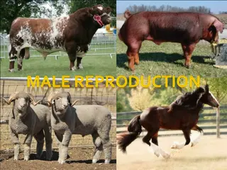
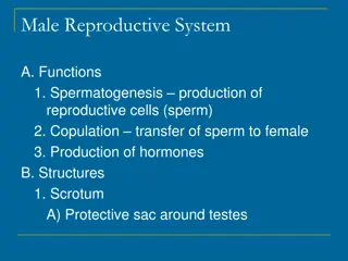
- The male reproduction system testes, scrotum, reproductive ducts, accessory sex glands, prepuce and penis - Production of - Spermatozoas Male sex hormone -Primary sex organs Testicles - Secondary sex organs (ductus system)- Epididymis, Vas deference, urethra, penis - Accessory sex glands Seminal vesicles, prostate and bulbo-urethral glands/cowpers gland
- SCROTUM SCROTUM . Cutaneous pouch . Encloses the testis . Testes fix by scrotal ligament . Scrotal hairs (except buck, ram, stallion and tom)
- Scrotal position Caudal to thighs Boar and Tom Between the thighs All others . Thermoregulation of testes . Sebaceous and sweat glands (Stallion, dog)
- SCROTUM LAYERS (outward to inward) 1. Epidermis 2. Dermis 3. Tunica dartos (fibroelastic CT & smooth muscles) 4. Tunica vaginalis communis/ reflexa (Extension of parietal peritoneum) 5. Vaginal process/cavity (space in between) 6. Tunica vaginalis propria (Extension of visceral peritoneum) 7. Tunica albuginea (fibrous layer with some smooth muscles) 8. Tunica vasculosa (layer with blood vessels)
- Tunica dartos - Form intertesticular septum - Stallion scrotum - short and non-pendulous - Testes in camel - non pendulous scrotum (in perineal region)
- The ductus system (in to out) : Seminiferous tubules Rete testes Efferent duct Caput epididymis Corpus epididymis Cauda epididymis Vas deferense Ampulla Pelvic urethra Penile urethra (in penis) . Exit
- SEMINIFEROUS TUBULES SEMINIFEROUS TUBULES Extension of tunica albuginea - Dividing testicular parenchyma into lobules (encloses seminiferous tubules) - Joins at mediastinum - Seminiferous tubules - Highly convoluted & Unbranched - Open at both ends - 75% of total testicular mass - They Have - Somatic sertoli cells/sustentacular/nurse - Sperm producing germ cells (spermatogonial germ cell line).
- The sertoli cells - Enrico Sertoli - The leydig cells - Franz Leydig. - Sertoli cells secretes mullerian inhibiting factor/hormone (MIH) (inhibite paramesonephric or mullerian duct) - The number of sertoli cells doesn t increase after puberty i.e. it limits the spermiogenesis or spermiogenic yield. - Sertoli cells form blood testes barrier just before the puberty to separate differentiating germ cells from blood circulation.
- Interstitial space (out of Sem. tub.) Leydig cells (interstitial cells) Blood vessels & Lymphatics - In ram leydig cells are found in clusters around blood vessels. - Length of seminiferous tubules : Boar 6 Km Bull 5 Km Ram 4 Km Dog 150 m Tom 25 m
- RETE TESTES RETE TESTES (in mediastinum) Seminiferous tubules Collecting tubules Rete testes Vas efferens - Lined by non secretory cuboidal epithelial cells
- TESTES TESTES (paired male gonads) - In scrotum at inguinal region - With help of tunics and spermatic cord - Covered with extra-abdominal tunic - Tunica vaginalis albugenia .vasculosa - Fluid in vaginal process - Movement of testis within scrotum - Plane of testes : Horizontal Stallion Vertical Cattle, ram, buck Oblique Boar, Dog, Tom
Testicular diameter Bull 5-9 cm Stallion 4-8 cm Ram and Buck 4-7 cm Boar 5-9 cm Dog 1-3 cm Tom 0.7 1.5 cm Shape of testes Oval stallion Elliptical boar Elongates oval bull, ram Round to oval Dog, Tom
- Size of testes varies throughout the year in seasonal breeder e.g. ram, stallion, camel - Weight of testis Bull 200-500 gm Ram and buck 200-400 gm Stallion 150-300 gm Boar 500-800 gm Dog 8-15 gm - In buffalo bull testes and scrotum are smaller and the penile sheath is less pendulous than cattle bull.
- Testes Endocrine Production of testosterone (By leydig cells .. LH/ICSH) Exocrine Spermatogenesis (FSH & testosterone) - Body weight .. positively correlated with testicular weight, testicular volume, total epididymal mass, total scrotal width - Castration (Removal of testicles) Reduces aggressive behavior of male animal Rreduces the boar taint
- Cremester Cremester muscle -Internal abdominal oblique muscle to tunica vaginalis propria muscle - Loosening & Raising of testes - In response to environmental temperature any abnormal stimulus is the - To maintain the testicular temperature - Retracting the testes within abdomen (Elephant, deer, and rabbit etc during their non breeding season)
- - Testes (in scrotum with help of spermatic cord) Spermatic cord comprises of . Internal spermatic artery . Spermatic veins (pampiniform plexus) . Lymphatic vessels . Autonomic nerves from renal and mesenteric plexus . Internal cremester muscle . Tunica vaginalis propria . Vas deferens - Mesorchium & mesepididymis
THANK YOU THANK YOU


")
")

 :")
 :")


")


")