Myoglobin and Hemoglobin: Oxygen-Binding Proteins in Evolutionary Demands
Myoglobin and hemoglobin are essential oxygen-binding proteins crucial for multicellular organisms and aerobic metabolic processes. Heme, with its iron atom, enables the reversible binding of oxygen to these proteins. The coordination bonds of iron in heme prevent oxidation by nitrogen atoms. The oxygen affinity of free heme molecules in water solution is influenced by the polypeptide environment. The structure of myoglobin, such as the sperm whale myoglobin, reveals insights into their functional properties and steric effects on ligand binding.

Download Presentation

Please find below an Image/Link to download the presentation.
The content on the website is provided AS IS for your information and personal use only. It may not be sold, licensed, or shared on other websites without obtaining consent from the author.If you encounter any issues during the download, it is possible that the publisher has removed the file from their server.
You are allowed to download the files provided on this website for personal or commercial use, subject to the condition that they are used lawfully. All files are the property of their respective owners.
The content on the website is provided AS IS for your information and personal use only. It may not be sold, licensed, or shared on other websites without obtaining consent from the author.
E N D
Presentation Transcript
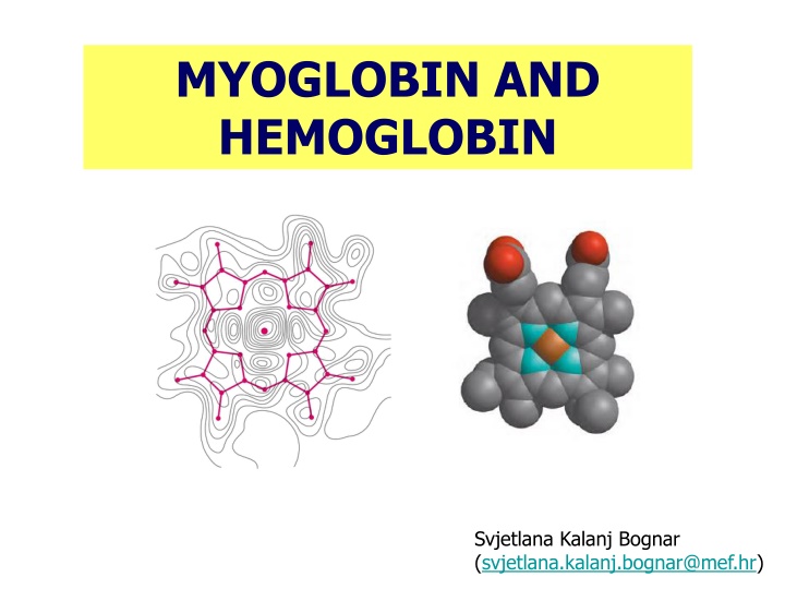
MYOGLOBIN AND HEMOGLOBIN Svjetlana Kalanj Bognar (svjetlana.kalanj.bognar@mef.hr)
Myoglobin and hemoglobin, oxygen-binding proteins evolutionary demands of multicellular organisms and aerobic metabolic processes oxygen solubility! Hemoglobin binds and transports oxygen in blood (erythrocytes). Myoglobin oxygen-binding protein, in muscle tissue, particularly abundant in the muscles of diving mammals (oxygen storage). 800_seals Killer%2520Whale
HEME IS THE PROSTHETIC GROUP ASSOCIATED WITH MYOGLOBIN AND HEMOGLOBIN. HEME CONTAINS IRON ATOM WHICH ENABLES REVERSIBLE BINDING OF OXYGEN. Prosthetic group is non-protein unit tightly attached to a complex protein structure, and required for specific function of that protein. 4 pyrrole rings are connected by methene bridges (=CH-); additional groups characteristic for heme porphyrin structure are 2vinyl groups (-CH=CH2), 2 propionate groups and 4 methyl groups. Heme group is present in different heme proteins. (a) protoporhyrin IX, organic ring structure containing four pyrrole rings; (b) Iron atom bound to the heme in ferrous (Fe2+) state which binds oxygen reversibly.
Iron atom in heme of myoglobin and hemoglobin has 6 coordination bonds: 4 to nitrogen atoms and 2 perpendicular to flat porphyrin ring system. What prevents the oxidation of Fe2+? Nitrogen atoms have electron-donating character and prevent conversion of heme iron from Fe2+to Fe3+ state.
What is the oxygen affinity of free heme molecules in a water solution? Polypeptide environment influences the function of a prosthetic group! Steric effects on binding of ligand to the heme of myoglobin. CO binds to free heme 20000 times better than oxygen, but 200 times better in myoglobin heme (steric hindrance and the role of distal histidine, CO cannot bind linearly!).
spermwhale Myoglobin structure was resolved by John Kendrew in 1957, using method of X-ray crystallography - for analysis, Kendrew used the myoglobin derived from muscular tissue of sperm whale. COO H NH2 MYOGLOBIN (Mr 16700; Mb) typical globin;153 amino acid residues, 78% of them in eight -helical segments (A-H) connected by bends.
What are the expected properties of amino acid residues in the interior and the outer part of the globular myoglobin molecule? Hydrophobic amino acids in the interior of the myoglobin structure (in yellow). distal histidine Almost all amino acid residues in the interior of myglobin molecule are nonpolar (leucine, valine, methionine, phenylalanine). Histidine residues are the only charged amino acid residues in the interior of myoglobin structure. proximal histidine
MYOGLOBIN IS OXYGEN STORAGE MOLECULE Graphical representation of ligand binding on the example of binding of oxygen to myoglobin hyperbolic curve. (partial pressure of O2 in the air above the solution expressed in kilopascals, kPa).
HEMOGLOBIN STRUCTURE was revealed in 1959 by Max Perutz and coworkers. Adult hemoglobin (HbA) Mr 64500, Hb 4 polypeptide chains 4 heme prosthethic groups 4 oxygen-binding sites 2 2 tetramer (2 chains - 141 amino acid residues; 2 chains - 146 amino acid residues) Oxygen saturation of Hb in arterial blood 96% Oxygen saturation of Hb in venous blood 64% In tissues, hemoglobin releases up to one third of the transported oxygen.
4 subunits (polypeptide chains) in hemoglobin structure are held together by interactions between hemoglobin subunits. Types of these interactions are hydrophobic, hydrogen bonds, ionic pairs. Ionic pairs are formed between between oppositely charged amino acid side chains in polypeptide chains of hemoglobin, and are involved in structural changes of hemoglobin during its oxygenation/de-oxygenation.
Structural changes of hemoglobin on binding oxygen: T state, deoxygenated Hb (tense, taut, low affinity state) and R state, oxygenated Hb (relaxed, high affinity state)/ T R transition
What is advantage of hemoglobin for its function as oxygen-transporter in blood red cells? Hb A binding curve of hemoglobin oxygen affinity: sigmoid binding curve reflects cooperative binding of oxygen which enables higher sensitivity of hemoglobin to small differences in pO2 between tissues and lungs. Mb 1. In myoglobin, hyperbolic binding curve shows insensitivity to small changes in the concentration of dissolved oxygen. 2. Quaternary structure of hemoglobin enables more sensitive response to small changes in ligand (oxygen) concentration.
Hemoglobin structure and function is an example of an allosteric protein: the binding of the ligand to one site affects the binding properties of another site on the same protein. Structural changes in a multisubunit protein undergoing cooperative binding to ligand ligand-binding induces higher affinity conformations of the protein.
Hemoglobin also transports H+ (40 % total H+)* and CO2 (15-20 % of CO2 formed in tissues)*, the end products of cellular respiration, to the lungs and kidneys. Hemoglobin binding sites for CO2are terminal NH2groups, and for H+side chains of amino acid residues in Hb structure, mostly histidine. carbaminohemoglobin *the remainder of the H+ is absorbed by plasma hydrogen carbonate buffer, and the remainder of CO2 is transported as dissolved HCO3- and CO2
The effect of pH and CO2 concentration on the binding and release of oxygen by hemoglobin is described as Bohr effect (Christian Bohr, 1904) When protonated, His HC3 forms ion pairs with Asp FG1, which stabilizes deoxyhemoglobin structure.
H+ produced in the reaction of carbamino-Hb formation contributes to Bohr effect!
lungs blood tissues Effects of pH on the binding of oxygen to hemoglobin. Both lower pH and increased concentration of CO2 decrease the Hb affinity for O2 binding.
Oxygen binding is regulated by 2,3-bisphosphoglycerate (BPG), which reduces the affinity of hemoglobin for oxygen. Binding of BPG to hemoglobin and stabilization of T (deoxyhemoglobin) state Negative charges of BPG interact with positively charged groups of amino acids in binding pocket which dissapears after oxygenation. BPG is the allosteric regulator of oxygen binding to hemoglobin.
Effect of BPG on the binding of oxygen to hemoglobin. (physiological adaptation to lower pO2 at higher altitudes, BPG concentration; also present in hypoxia, and certain respiratory diseases)
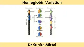
Fetal hemoglobin (HbF) does not bind 2,3-BPG as well as adult hemoglobin. Fetal Hb (90% saturation) Fetal hemoglobin structure is 2 2 tetramer. Fetal hemoglobin needs to have greater affinity for oxygen. File:Postnatal genetics en.svg
COHb in blood and CO in the air Oxygen-binding curves compared Intoxication with carbon monoxide -formation of carboxyhemoglobin: 10% of COHb no symptoms; 15% of COHb mild headaches; 20-30% of COHb severe headaches, nausea, confusion, disorientation, visual disturbances; 30-50% of COHb, neurological symptoms; 50% of COHb, loss of consciousness and coma, respiratory failure; death above 60% of COHb)
CLINICAL APPLICATIONS ALTERATIONS IN STRUCTURE OF HEMOGLOBIN, LEADING TO DISORDERED FUNCTION OF HEMOGLOBIN Sickle cell anemia molecular disease of hemoglobin First case described in 1904 J. Herrick, Chicago physician REPLACEMENT OF ONE AMINO ACID IN HEMOGLOBIN STRUCTURE LEADS TO DISEASE! Linus Pauling described altered electrophoretical properties of HbS. Vernon Ingram sequenced altered HbS glutamate at 6th position of beta chain is replaced with valine.
Methemoglobinemia - Hemoglobin M 1. Congenital - example of the mutation in the active site Replacement of proximal or distal histidine with tyrosine causes stabilization of the heme in Fe3+ form in which binding of oxygen is not possible! Change may occur in both types of polypeptide chains (alfa or beta). Mutant hemoglobin is called methemoglobin (HbM). Patients are cyanotic, only heterozygous carriers have been detected (homozygosity would lead to death). 2. Acquired - caused by exposure to certain medications GLYCATED HEMOGLOBIN Determined HbA1clevel is a relatively accurate measure of the amount of glucose in the blood and the length of time the concentration of blood glucose has been elevated. (Glucose reacts nonenzymatically with amino groups of amino acid residues in Hb -chains.)
SUMMARY 1. Myoglobin and hemoglobin contain heme prosthetic group containing iron atom in its ferrous state (Fe2+)which binds oxygen reversibly. 2. Normal adult hemoglobin has four heme-containing subunits, similar in structure to myoglobin. 3. Oxygen binding to hemoglobin is allosteric and cooperative. As oxygen binds to one binding site, hemoglobin undergoes conformational changes which affect other binding sites. Conformational changes between T and R state result in cooperative binding. 4. Hemoglobin binds H+ and CO2 (Bohr effect). Oxygen binding to hemoglobin is also modulated by 2,3-bisphosphoglycerate. Literature: 1. D.L. Nelson i M.M. Cox: Lehninger Principles of Biochemistry, 5thedition, Worth Publishers, USA, 2008. 2. J.M. Berg, J.L. Tymoczko i L. Stryer: Biochemistry, 7th edition, W.H. Freeman and Company, USA, 2012 Link to website Medical Biochemistry Page - Myoglobin and hemoglobin
REVIEW QUESTIONS 1. The muscle protein myoglobin and the erythrocyte protein hemoglobin are both oxygen transport proteins. Describe the structural features that allow these molecules to perform their separate functions. 2. Fetal hemoglobin (HbF) binds to BPG to a lesser extent than does adult hemoglobin (HbA). Why do you think HbF has a greater affinity for oxygen than does maternal hemoglobin?
ANSWERS TO REVIEW QUESTIONS 1. The structural differences between two proteins underlie their different functions. Myoglobin contains one polypeptide chain and one heme as a prosthethic group, while hemoglobin consists of 4 polypeptide chains and 4 hemes. Myoglobin has high affinity for oxygen binding which makes it a perfect oxygene storage protein. Hemoglobin acts as an allosteric protein, as there is cooperative binding of its ligand, oxygen, to iron (Fe2+) in heme. Hemoglobin is more sensitive to small changes in partial pressure of oxygen, and is perfect transporter of oxygen in blood. (Remind that the binding of oxygen is called oxygenation. This should not be confused with oxidation, in which iron loses electron and acquires Fe3+oxidation state. If oxidation of heme iron occurs, which is related to certain disorders, there is no binding of oxygen at all!) 2. Fetal hemoglobin (HbF) has different structure than adult hemoglobin (HbA) it contains 2 and 2 chains, and due to this structural difference binding of 2,3 BPG is not possible for fetal hemoglobin. Thus, there is no effect of 2,3 BPG which decreases affinity of hemoglobin to oxygen binding. Physiologically, this structural feature of fetal hemoglobin and its higher affinity for oxygen than adult hemoglobin enables efficient fetal utilization of oxygen from maternal circulation.


 in hemoglobin structure")


* and")


")