Odontogenic Cysts and Tumors
Odontogenic cysts and tumors originate from the odontogenic apparatus and can be classified into various types including inflammatory and developmental. Understanding the characteristics and differentiation of these lesions is crucial for accurate diagnosis and treatment planning. From periapical cysts to odontogenic tumors, each entity presents unique features that impact patient management and outcomes.

Download Presentation

Please find below an Image/Link to download the presentation.
The content on the website is provided AS IS for your information and personal use only. It may not be sold, licensed, or shared on other websites without obtaining consent from the author.If you encounter any issues during the download, it is possible that the publisher has removed the file from their server.
You are allowed to download the files provided on this website for personal or commercial use, subject to the condition that they are used lawfully. All files are the property of their respective owners.
The content on the website is provided AS IS for your information and personal use only. It may not be sold, licensed, or shared on other websites without obtaining consent from the author.
E N D
Presentation Transcript
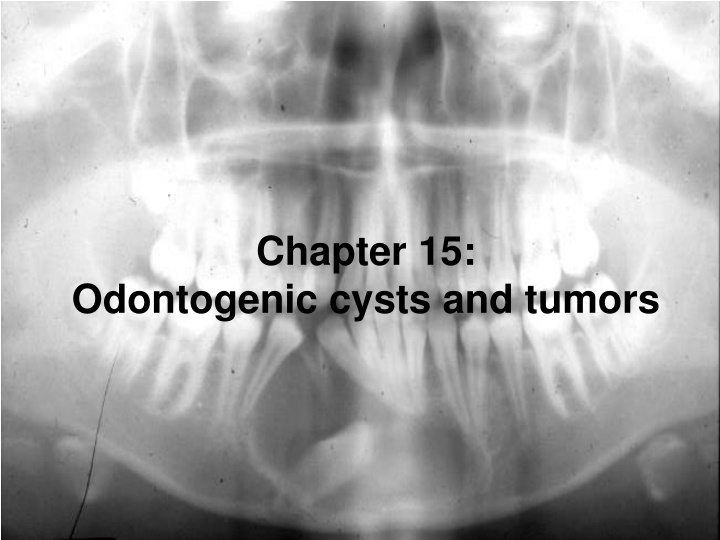
Chapter 15: Odontogenic cysts and tumors
Overview: Odontogenic cysts & tumors arise from the odontogenic apparatus. The odontogenic apparatus consists of: Epithelium: Remnants of dental lamina Reduced enamel epithelium Odontogenic rests Lining of odontogenic cysts Basal cell layer of oral mucosa Ectomesenchyme: Dental papilla Q: What is a cyst? A: An abnormal space within tissue lined by epithelium. Q: Name some cysts that are not really cysts: A: Aneurysmal bone cyst, Stafne bone cyst, Traumatic bone cyst, Simple bone cyst, Eruption cyst Q: Why are they not cysts? A: No epithelial lining!
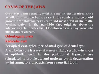
The Cysts and Tumors of Chapter 15: Odontogenic cysts: Inflammatory: Periapical (radicular) cyst Residual periapical (radicular) cyst Buccal bifurcation cyst (usually first molars) Paradental cysts (partially erupted third molars Developmental: Dentigerous cyst Odontogenic keratocyst (KOT) Orthokeratinized odontogenic cyst Gingival (alveolar) cyst of the newborn Gingival cyst of the adult Lateral periodontal cyst Calcifying odontogenic (Gorlin) cyst Glandular odontogenic cyst Eruption cyst Odontogenic Tumors: Epithelial Tumors: Ameloblastoma Adenomatoid odontogenic tumor Calcifying epithelial odontogenic tumor (Pindborg tumor) Squamous odontogenic tumor Clear cell odontogenic carcinoma Ectomesenchymal Tumors: Odontogenic myxoma Granular cell odontogenic tumor Central odontogenic fibroma Cementoblastoma Mixed Odontogenic Tumors: Odontoma Compound Complex Ameloblastic fibroma Ameloblastic fibro-odontoma Ameloblastic fibrosarcoma Odontoameloblastoma
Periapical cyst or granuloma (chronic localized osteitis) Impossible to tell radiographically which one it is only histologically, so you must include both in your differential diagnosis. Q: Why does a periapical cyst form instead of just a granuloma? A: If by chance there are Rests of Malassez in the area of inflammation. The rest cells proliferate due to the inflammation The ball of cells gets too large, cells in the center die, center then has a higher protein concentration, water rushes in to equalize the osmotic pressure. Osmotic pressure can continue to grow the cyst independent of the inflammation. Other unilocular radiolucencies located periapically: (early) periapical cemento-osseous dysplasia teeth are vital Dentin dysplasia type I teeth are vital, multiple radiolucencies With a periapical cyst or granuloma, the tooth is NON-VITAL Take a vitality test!! Tx for a non-vital tooth is root canal. Must biopsy a radiolucent lesion beneath a vital tooth.
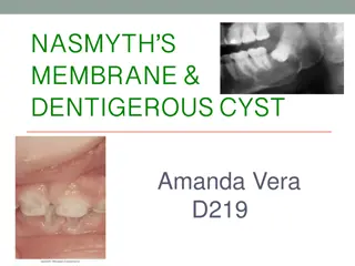
Dentigerous cyst Radiolucency associated with an unerupted tooth encloses the crown of the unerupted tooth and is attached at the CEJ Most common developmental odontogenic cyst Should be the first differential diagnosis for any radiolucency associated with an unerupted tooth Others: Odontogenic Keratocyst (KOT), Ameloblastoma
043bb 042bb (Vital teeth) 039bb 041bb
Odontogenic Keratocyst (Keratocystic Odontogenic Tumor) Can be in the location of ANY other type of odontogenic cyst or can be isolated in the jaws! a benign uni-or multicystic, intraosseous tumor of odontogenic origin lining is parakeratinized stratified squamous epithelium potential aggressive, infiltrative behavior solitary or multiple (multiple usually related to Gorlin syndrome) Three important things associated with this diagnosis: 1. High recurrence rate (up to 60%) 2. Highly aggressive (now considered by W.H.O. to be an odontogenic tumor) 3. Relation to Gorlin syndrome Arises from the dental lamina or its remnants PTCH gene is a significant factor in the development of KOT
Nevoid Basal Cell Carcinoma Syndrome (Gorlin Syndrome) Multiple basal cell carcinomas Multiple jaw cysts (odontogenic keratocysts) Numerous bone abnormalities including bifid ribs, intracranial calcification, vertebral anomalies PTCH gene has been mapped to chromosome 9q22.3 - site of Gorlin Syndrome Anyone with multiple KOTs should be tested for Gorlin Syndrome
062bb 061bb
Lateral Periodontal Cyst The lateral periodontal cyst is generally quite small and well demarcated. It occurs most frequently in the mandibular bicuspid area adjacent to vital teeth. Radiolucencies are generally small and ovoid Derived from remnants of the dental lamina Tx: conservative enucleation Considered to be the intrabony counterpart to the Adult Gingival Cyst
Calcifying Cystic Odontogenic Tumor (Gorlin s cyst) Uncommon lesion that demonstrates considerable histopathologic diversity and variable clinical behavior Can be unilocular or multilocular, can be associated with an unerupted tooth Tx: simple surgical excision, prognosis is usually good Ghost cells calcify
137bb 135big 133bb