Explore the different types of chronic liver diseases, including hepatitis and cirrhosis, along with information on diagnosis, treatment, and prevention. Learn about autoimmune liver diseases, common disorders like NASH and alcoholic liver disease, and the role of aminotransferases in assessing liver health.
Please find below an Image/Link to download the presentation.
The content on the website is provided AS IS for your information and personal use only. It may not be sold, licensed, or shared on other websites without obtaining consent from the author. If you encounter any issues during the download, it is possible that the publisher has removed the file from their server.
You are allowed to download the files provided on this website for personal or commercial use, subject to the condition that they are used lawfully. All files are the property of their respective owners.
The content on the website is provided AS IS for your information and personal use only. It may not be sold, licensed, or shared on other websites without obtaining consent from the author.
E N D
Presentation Transcript
Chronic Chronic hepatitis/ hepatitis/hepatopathy Liver Liver cirrhosis cirrhosis hepatopathy Department of Internal Medicine Second Faculty of Medicine, Charles University and Motol University Hospital
Chronic Chronic liver liver disorders disorders early diagnosis and treatment to prevent liver fibrosis and cirrhosis
Types Types of of liver liver diseases diseases sorted sorted by etiology by etiology hepatocytes biliary tree INFECTIOUS TOXIC METABOLIC (impairment of metabolism and/or of transport) AUTOIMMUNE TUMOROUS VASCULAR
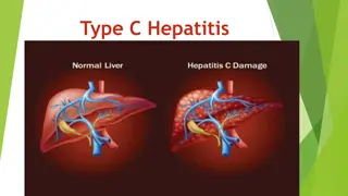
The most common chronic hepatopathy alcoholic liver disorders NASH chronic hepatitis C, chronic hepatitis B, B+D autoimune hepatitis cholestatic liver disorders PBC primary bilary cholangitis PSC primary sclerosing cholangitis Wilson disease hereditary hemochromatosis deficit alfa-1 antitrypsinu
AUTOIMMUNE LIVER DISEASE hepatocytes biliary tree = cholestatic disorders autoimmune hepatitis (AIH) - typ I, II, III PBC (primary biliary cholangitis) PSC (primary sclerosing cholangitis) IgG4 associated cholangitis overlap syndromes AIH/PBC, AIH/PSC, AIH/chronic hepatitis C Target cells for autoimmune reaction are: hepatocytes (in AIH) cholangiocytes of proximal bile ducts (in PBC) of distal bile ducts (in PSC and IgG4 ass.cholangitis)
Autoimmune Autoimmune hepatitis ( hepatitis (AIH) AIH) AIH 4x frequent in F AIH relatively common in childhood (typ II) and young women typically: chronic hepatitis (2 % of all chron. hep.) progressive necro-inflammatory process progression to fibrosis cirrhosis BUT: 1/3 of cases: acute hepatitis (icterus, ALT a AST), also fulminant it could be also random dg.
Aminotransferases ~ hepatocyte integrity ~ histological activity of hepatic inflammation AST (up to 0,92 kat/l) ALT (up to 0,73 kat/l) hepatocyte cytoplasm hepatocyte cytoplasm mitochondria skeletal muscle, cardiac muscle (lung, pancreas, spleen, kidney)
AIH AIH immunological immunological diagnostis serum protein ELFO: Ig (polyclonal) ANA (anti-nuclear Ab) ASMA (anti-smooth muscle Ab) AAA (anti-actin Ab) SLA (soluble liver antigen Ab) anti-LKM (anti-liver kidney microsomal antigen 1) - against proteins of ER in hepatocytes and proxim.tub. + in DILI (drug inducer liver injury) and chron. HCV anti-ASGPR (anti-asialoglykoprotein receptor Ab) + in PBC, PSC, and other hepatitis anti-LC1 (anti-liver cytosol 1 Ab) the most specific in ?dg anti-LP (anti-liver pancreatic antigen) diagnostis
AIH AIH AIT type I - associace with other AI disease: pernicious anemia, AIHA, autoimmune thyroiditis AIH type I: + AMA, + ASMA, (pANCA, cANCA, anti-dsDNA) younger women, tendency to progress to cirrhosis AIH type II: + anti-LKM subtype II b: + anti HCV (HCV-RNA) AIH typ III: + SLA (clinically like typ I)
AIH AIH - - histology histology 1/ characteristic (not pathognomonic dif. dg. viral or toxic hepatitis) : in portal tracts: infiltrate of lymfocytes and plasma cells) (interface hepatitis) piecemeal necrosis bridging necrosis fibrotisation grading (inflammation) stading (fibrotisation) bile ducts intact bile duct
If dg. ? ? scoring systems (basis) + point: F, ALP/AST < 1,5 Ig 2x ULN autoantibodies histology: periportal inflammation (interface hepatitis) infiltrate of lymfocytes and plasma cells other AI disease - point: M; ALP/AST 3 alcohol consumption 60g/d hepatotoxic drugs active viral infection AMA
Therapy Therapy of of AIH AIH immunosuppressive drugs glucocortikoids: prednison 40-60 mg/d after the efect of therapy (AST, ALT activity) 20 mg/d azathioprine 50-100 mg/d other immunosuppressants (cyclosporin, tacrolimus, mycofenolate mofetil) relapsing; HCC is rare Liver Tx (fulminant AIH, cirrhosis)
AUTOIMMUNE LIVER DISEASE hepatocytes biliary tree = cholestatic liver disorders autoimmune hepatitis (AIH) - typ I, II, III - IgG4 hepatitis PBC (primary biliary cholangitis) AIC (autoimmune cholangitis) PSC (primary sclerosing cholangitis) IgG4 associated cholangitis overlap syndromes AIH/PBC, AIH/PSC, AIH/chronic hepatitis C
Cholestatic Cholestatic liver liver diseases diseases chronic cholestasis more than 6 month ALP 1,5x ULN GGT 3x ULN
PBC PBC Histology staging: 1. lymphocytic infiltrate (florid ductal lesion) in portal tracts with granulomatous desctruction of portal bile ducts ductopenia (vanishing bile duct syndrome) acute and chronic cholestatic changes (bilirubinostasis, ) portal/periportal inflammation 2. marginal proliferation of small bile ducts 3. fibrosis (portal periportal 4. cirrhosis HCC Clinical features: pruritus jaundice xantomas (retained cholesterol) steatorhea (vit D deficiency )
PSC PSC periductal inflammation and obliterative fibrosis of extrahepatic and intrahepatic bile ducts segmental stenosis dilation of preserved segments Histology: onion skin fibrosis
PBC: microscopic bile ducts v 90-95 % in F pruritus (anicteric cholestasis) precedes dg. of PBC icterus unfauvorable sign PSC: macroscopic bile ducts M:F = 2:1 in 80 % precedes UC (less mC) icterus, cholangoit dy hnisav secondary biliary cirrhosis and cholangiocarcinoma (Ca 19-9) (ANA, ASMA) ERCP, MRCP (liver biopsy not dg.) IgM, +AMA (M2), ev. ANA abdominal US for dif. dg. histology in dg.
Therapy Therapy of of PBC PBC and and PSC PSC UDCA 15 20 mg/kg daily, improves/not improves survival UDCA + glucocorticoid (budesonid) 2nd linie: obeticholic acid (modificated bile acid) ATB (second. cholangitis), therapeutical ERCP: baloon dilation and stenting Therapy of pruritus antihistaminics bile acid sequestrants cholestyramine (bounds BA in GIT) rifampicin (inhibition of E-H circulation: inhibition of BA uptake by hepatocytes) nasobiliary drainage Th of osteoporosis (deficiency of vit. A,D,E,K) Liver Tx
Hereditary Hereditary hemochromatosis hemochromatosis Basic knowledge: iron metabolism: total body iron content regulated by intestinal absorption (hepcidin and other proteins) hemochromatosis = heritable (AR), caused by excessive Fe intestinal absorption hemosiderosis = disorder associated with parenteral Fe administration (repetitive transfusions), ineffective erythropoiesis or with chronic liver disease
Hepcidin Hepcidin iron regulatory hormone - secreted by hepatocytes - blockes the expression of ferroportin (Fe efflux chanel on intestinal epitelium and on macrofages) - hepcidin lowers plasma iron hepcidin deficiency Fe overload - Bacterial sepsis: IL-6 stimulates sekretion of hepcidin: S-Fe ( intest.absorption + retention in macrofages) reproduction of bakteria
Other Other proteins proteins involved involved in in Fe Fe metabolism metabolism e.g. HFE (high Fe), hemojuvelin (HJV), transferrin receptor 2 modulate hepcidin levels
Hereditary Hereditary hemochromatosis hemochromatosis - excessive Fe intestinal absorption - excessive Fe accumulation in the parenchymal cells of various organ: - Fe toxicity (free radical formation, lipid peroxidation) tissue damage liver: sideronecrosis of hepatocytes, activation of stellate cells fibrogenesis cirrhosis pancreas: diabetes melllitus (bronze) skin: hyperpigmentation ( melanin) heart: cardiomyopathy (arrhythmias, heart failure) joints: artropathy (Fe accumulation in synovial cells) gonad: atrophy - hypogonadism
Hereditary Hereditary hemochromatosis hemochromatosis- - diagnosis diagnosis transferin saturation > 50-60 % (n: 16-45 %) transferin (n: 2,0-3,6 g/l) feritin (n: 10-290 g/l) > 1000 g/l CAVE: feritin = acute phase reactant inflammation, mlg. liver biopsy: confirm clinical impression + staging (fibrosis) Fe in dry weight of liver (n: 35 mol/g) the hepatic iron index: > 1,9 Fe in dry weight/atomic weight of Fe/age
Hereditary Hereditary hemochromatosis hemochromatosis clinical clinical features features Fe accumulation is life-long injury caused by Fe is gradual symptoms after age of 40 years M/F = 6/1 (physiological Fe loss in F: menstruation, pregnancy) fully developed: micronodular ci + DM + skin pigm. death: complication of cirrhosis (incl. HCC) cardiac involvement Th: regular phlebotomies: 500 ml (250mg Fe) 1-2x weekly to feritin < 50 ng/ml (chelating agents: desfreroxamin, deferipron, ..) !!! early diagnosis (20 yo) = normal life expectancy screening of genetic probands
Hereditary Hereditary hemochromatosis hemochromatosis- - genetics genetics the most common inherited disease in individuals of Northern European ancestry (highest in Celtic origin) AR; HFE gene (short arm of chromos. 6) an estimated carrier rate of 1 in 10 frequency homozygosity ~ 1:200-400 but low penetrance (genetic background does not always lead to disease) molecular genetic tests HFEhemochromatosis HFE mutation: C282Y (Cys282Tyr), H63D (His63Asp), S65C (Ser65Cys) - homozygot - compound heterozygot - in 5 % new mutation
Wilson Wilson disease disease AR, mutations of the ATP7B gene (canalicular copper-transporting ATPase) Cu absorption and delivery to the liver is normal, but: reduced Cu excretion into the bile and impaired incorporation of Cu into ceruloplasmin Cu accumulation in liver hepatic injury spillover of Cu (non-ceruloplasmin-bound) into the circulation hemolysis accumulation and injury in brain, cornea = Clinical features: A/CH liver disease neuropsychiatric changes young women - haemolytic anemia
Wilson Wilson disease disease liver liver damage damage minor to .severe acute (fulminant ~ massive necrosis) hepatitis or chronic hepatitis cirrhosis fatty change
Wilson Wilson disease disease clinical clinical features features age of onset (variable) before age 40 years hepatic involvement hemolytic anemie (young women) neuropsychiatric disorders mild behavioral changes frank psychosis Parkinson-like symptoms (Cu in basal ganglia)
Wilson Wilson disease disease diagnosis diagnosis ( of serum ceruloplasmin, serum Cu) urinary Cu excretion (penicillamine test), liver biopsy content of Cu in dry liver tissue staging family screening: mutation of the ATP7B gene
Wilson Wilson disease disease therapy therapy avoid excess in copper-containing foods (mushrooms, nuts, chocolate, dried fruit, liver, shellfish) excretion of copper in the urine: p.o. chelators: penicillamine (SE: drug-induced lupus, myasthenia), trientine absorption of copper from the intestine: p.o. zinc acetate in rare (severe neurological) cases: i.m. dimercaprol
1 1- -antitrypsin antitrypsin deficiency deficiency 1-AT - serum protease inhibitor (leukocyte elastase): -synthesized byl hepatocytes - AR - risk allele homozygotes (PiZZ): 1-AT level below 10% of normal in the liver liver hepatocytes - polymerization of structurally defective protein - retention in the ER ER stress response activation of caspases mitochondrial damage > inflammation, fibrosis, PAS+ cytoplasmic globules systemic effect of 1-antitrypsin deficiency = protease activity (neutrophil elastase disrupts connective tissue) - lung emphysematous damage (pulmonary emphysema, COPD) ! smoking - panniculitis, arterial aneurysms, pancreatitis,
1 1- -antitrypsin antitrypsin deficiency deficiency Diagnostics: reduced levels of 1-AT (15% of normal) genetic testing - Pi allele M, S, Z, null (10% of neonatal hepatitis) hepatology -neonatal hepatitis with cholestatic icterus -adulhood: cholestasis, hepatitis, cirhosis 2-3 % HCC -histology: PAS + globules, cholestasis Treatment: - lung-affected (not liver-affected) patients may receive i.v. infusions of alpha-1 antitrypsin - studies: recombinant and inhaled forms of A1AT
Liver Liver fibrogenesis fibrogenesis clinical clinical issue issue It is reversible Methods of testing of liver fibrosis products of stellate cells released into the blood: (hyaluronic acid, alfa-2-microglobulin, collagenous markers, matrix metalloproteinases) Indexes based on routine lab. tests: Measurement based on imaging methods: fibroscan, US-/MR- elastography 36
Spontaneous Spontaneous bacterial bacterial peritonitis (SBP) peritonitis (SBP) Bacterial translocation into ascites Symptomatology: pure Dg puncture of ascites: Neutrofils 0,25 x 10 /l or WBCs 0,5 x 10 /l and/or + cultivation Th: cefalosporins 3. gen (G-) cefotaxim 2,0 g i.v. 2xd at least 5 days (ceftazidim, ceftriaxon) SBP aquired in hospital (G+ cocci): karbapenems or piperacilin/tazobactam Secondary prevention after episode of SBP: norfloxacin 400 mg p.o. 1x d or better: rifamixin 400 mg 3x d to the vanishing of ascites to the liver Tx
Hepatorenal Hepatorenal syndrome syndrome Functional renal failure in patients with liver cirrhosis and portal HT: vasoconstriction in renal arteries GF retention of water and Na Type 1 quickly progredient During 2 weeks twice incresed S-creatinine (at least 221 mol/l) Th: terlipresin, albumin 20% 100-200 ml/d hemodialysis Type 2 chronic , associated with refractory ascites and liver insufficiency Th: 1/ treatment of ascites: paracenthesis + albumin 2/ terlipresin 3/ HD, diuretics ? however not decrease intravascular volume
Examination Examination in evaluation evaluation of of risk in compensated compensated liver risk of of decompensation decompensation liver cirrhosis cirrhosis Define etiology of liver cirrhosis treatment according to the etiology Gastroscopy ? Presence of varices and primary prophylaxis of bleeding Ultrasonography: screening of HCC Non-invasive assesment of liver stiffness Markers of viral hepatitis A and B Vaccination for prevention of acute infection
Medications Medications contributing contributing to liver liver cirrhosis to decompensation decompensation of of cirrhosis Non-steroidal antiinflammatory drugs Decrease effect of diuretics, leed to renal vasoconstriction, risk of hepatorenal syndrome Vasodilators Deepen circulatory dysfunction (hyperkinetic circulation) and risk of hepatorenal syndrome Non-selective betablockers and carvedilol Not use when connected with decrease of BP
Liver Liver transplantation transplantation - - indication indication First decompensation Icterus New ascites After variceal bleeding First signs of liver encephalopathy Hyponatremia Hepatorenal or hepatopulmonal syndrome Acute on chronic liver failure Child-Pugh B or MELD 14-15 points Hepatocellular carcinoma (Milan criteria)
Take Take- -home home message message Cirrhosis is a heterogeneous and dynamic disease With appropriate approaches there can be: Prevention/delay of progression Regression of cirrhosis to earlier stages The approach to each stage will be different and depend on the prevailing pathophysiological mechanism(s) In end-stage liver cirrhosis timing of liver transplantation


")