Pulmonary Embolism: Causes, Risks, and Symptoms
An overview of pulmonary embolism, an obstruction in the pulmonary artery, including types, mortality rates, risk factors, and common symptoms. Explore the anatomy and clinical presentation of acute PE. Learn about the major and minor risk factors of venous thromboembolism. Recognize the history and presentation with common symptoms and worrying signs to watch for.

Download Presentation

Please find below an Image/Link to download the presentation.
The content on the website is provided AS IS for your information and personal use only. It may not be sold, licensed, or shared on other websites without obtaining consent from the author.If you encounter any issues during the download, it is possible that the publisher has removed the file from their server.
You are allowed to download the files provided on this website for personal or commercial use, subject to the condition that they are used lawfully. All files are the property of their respective owners.
The content on the website is provided AS IS for your information and personal use only. It may not be sold, licensed, or shared on other websites without obtaining consent from the author.
E N D
Presentation Transcript
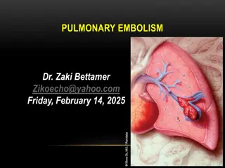
Pulmonary Embolism Chad Eastwood ST3 Respiratory
General: An obstruction pulmonary artery or one of its branches by Material that originates somewhere else in the body Main types include Thrombus, fat, air and amniotic fluid. After coronary artery disease and stroke, acute PE ranks third among the most common types of cardiovascular diseases. PE occur at ages 60 -70 years, autopsy data show the highest incidence among individuals 70 to 80 years of age.
Mortality (Exp Clin Cardiol. 2013 Spring; 18(2): 129138) Clinical presentation of acute pulmonary embolism Mortality rate Unselected population 11.4% at 2 weeks, 17.4% at 3 months Massive pulmonary embolism Overall 18% to 65% Treated Approximately 20% With cardiogenic shock 25% to 30% With resuscitation 65% Submassive pulmonary embolism 5% to 25% Pulmonary embolism with mobile thrombi in right- heart chambers As high as 27% Small pulmonary embolism Up to 1%
Risk factors of venous thromboembolism(British Thoracic Society, 2003) Major risk factors (RR = 5 to 20) Minor risk factors (RR = 2 to 4) Postoperative states: Major abdominal/pelvic surgery, hip/knee joint replacement, postoperative intensive care Obstetrics: Late pregnancy, Caesarian section, puerperium Lower limb affections: Fractures, extensive varicosities Malignancies: Abdominal/pelvic, advanced/metastatic stage Limited mobility: Hospitalization, geriatric care Miscellaneous: History of previous venous thromboembolism Cardiovascular: Congenital heart disease, heart failure, hypertension, superficial venous thrombosis, central venous catheter Humoral: Estrogen use: oral contraception, hormone replacement therapy Miscellaneous: Chronic obstructive lung disease, neurological impairment, latent malignancy, thrombotic defects, long-distance travel in the sitting position, obesity Other: Inflammatory bowel disease, nephrotic syndrome, chronic dialysis, myeloproliferative disease, paroxysmal nocturnal hemoglobinuria
History and presentation: Common symptoms include: SOB, Chest pain, swollen(unilateral) painful limb, cough, haemoptysis or exertional dyspnoea. Worrying symptoms: presyncope or syncope or impending sense of doom or sweating
Useful clues in the history: Past medical/surgical hx: previous DVT/ PE, clotting disorders, recent surgery Family hx: clotting disorders ie sickle cell Medications: OCP Social hx: prolonged immobility ie recent flight
Observations: HR: tachycardia (bradycardia is a worrying feature) BP (low in massive PE) o2 (Reduced) RR (tachypnoeic) Temperature: usually normal
Investigations: Bedside Bedside:
Bloods: FBC: raised WCC often or normal U+E: help dose anticoagulation CRP: may be normal or raised D-Dimer: (Only perform if suspicion of PE; High sensitivity low specificity) BNP: indicating RSHF Troponin: cardiac myocyte damage
Treatment of PE Depends on type of PE and clinical situation 1) Massive PE: (Hypotension SBP<90 for 15 mins and haemodynamically unstable) - Thrombolysis (alteplase 10mg bolus and 90mg over 2 hour) 2) Sub-massive PE: (normal BP, RHS and large clot burden, elevated troponin and raised bnp) Anticoagulation oral or sc Dosing is important and depends on weight and renal function Duration of treatment Complications Contraindications Counselling Monitoring
Duration: (Thorax 2003:58;470-484) Provoked PE with Temporary risk factor 3 months First idiopathic PE 3 months Other categories of PE 6 months Previous PE or DVT Long term
Assessment to discharge: Pulmonary embolism servity index( PESI)


: 129–138)")


")
")


")