Understanding Respiration and Muscles Involved in Breathing
Explore the process of respiration, including lung inflation, gas exchange in the alveoli, and the key muscles of breathing like the diaphragm and intercostal muscles. Gain insights into the role of these muscles in expanding the chest during inspiration and learn how the diaphragm contributes to separating the thoracic and abdominal cavities.

Download Presentation

Please find below an Image/Link to download the presentation.
The content on the website is provided AS IS for your information and personal use only. It may not be sold, licensed, or shared on other websites without obtaining consent from the author. If you encounter any issues during the download, it is possible that the publisher has removed the file from their server.
You are allowed to download the files provided on this website for personal or commercial use, subject to the condition that they are used lawfully. All files are the property of their respective owners.
The content on the website is provided AS IS for your information and personal use only. It may not be sold, licensed, or shared on other websites without obtaining consent from the author.
E N D
Presentation Transcript
RESPIRATION Done by: Mrs. Mercy Deva Priya Asst.Professor .Dept of MHN
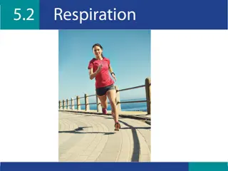
RESPIRATION Inflation and deflation of the lungs occurring with each breath ensures that regular exchange of gases takes place between the alveoli and the external air.
Muscles of respiration The expansion of the chest during inspiration occurs as a result of muscular activity, partly voluntary and partly involuntary. The main muscles of respiration in normal quiet breathing are the intercostal muscles and the diaphragm. During difficult or deep breathing they are assisted by the muscles of the neck, shoulders and abdomen.
Intercostal muscles There are 11 pairs of inter-costal muscles that occupy the spaces between the 12 pairs of ribs. They are arranged in two layers, the external and internal inter-costal muscles
The external inter-costal muscle fibres. These extend in a downwards and forwards direction from the lower border of the rib above to the upper border of the rib below. The internal inter-costal muscle fibres. These extend in a downwards and backwards direction from the lower border of the rib above to the upper border of the rib below, crossing the external inter-costal muscle fibres at right angles.
The first rib is fixed. Therefore, when the inter- costal muscles contract they pull all the other ribs towards the first rib. Because of the shape of the ribs they move outwards when pulled upwards. In this way the thoracic cavity is enlarged antero-posteriorly and laterally. The inter-costal muscles are stimulated to contract by the inter-costal nerves.
Diaphragm The diaphragm is a dome-shaped structure separating the thoracic cavities. It forms the floor of the thoracic cavity and the roof of the abdominal cavity and consists of a central tendon from which muscle fibres radiate to be attached to the lower ribs and sternum and to the vertebral column by two crura. and abdominal
When it contracts, its muscle fibres shorten and the central tendon is pulled downwards to the level of the 9th thoracic vertebra, enlarging the thoracic cavity in length. This decreases pressure in the thoracic cavity and increases it in the abdominal and pelvic cavities. The diaphragm is supplied by the phrenic nerves.
The inter-costal muscles and the diaphragm contract simultaneously enlargement of the thoracic cavity in all directions, that is from back to front, side to side and top to bottom. ensuring the
Cycle of respiration This occurs 12 to 15 times per minute and consists of three phases: inspiration expiration pause.
Inspiration-is as active process When the capacity of the thoracic cavity is increased by simultaneous contraction of the intercostal muscles and the diaphragm, the parietal pleura moves with the walls of the thorax and the diaphragm. This reduces the pressure in the pleural cavity to a level considerably lower than atmospheric pressure. The visceral pleura follows the parietal pleura pulling the lung with it. This stretches the lungs and the pressure within the alveoli and in the air passages falls, drawing air into the lungs in an attempt to equalise the atmospheric and alveolar air pressures.
Expiration Relaxation of the inter-costal muscles and the diaphragm results in downward and inward movement of the rib cage and elastic recoil of the lungs. As this occurs, pressure inside the lungs exceeds that in the atmosphere and therefore air is expelled from the respiratory tract. The lungs still contain some air and are prevented from complete collapse by the intact pleura. This process is passive as it does not require the expenditure of energy. After expiration, there is a pause before the next cycle