When a bacterial cell is deposited, it divides and forms colonies with distinct cultural characteristics. Understanding growth patterns, pigmentation, and optical characteristics on nutrient broth and slants aids in identification. Explore the characteristics of bacteria and fungi to differentiate between them based on colony morphology and growth behavior.
Please find below an Image/Link to download the presentation.
The content on the website is provided AS IS for your information and personal use only. It may not be sold, licensed, or shared on other websites without obtaining consent from the author. If you encounter any issues during the download, it is possible that the publisher has removed the file from their server.
You are allowed to download the files provided on this website for personal or commercial use, subject to the condition that they are used lawfully. All files are the property of their respective owners.
The content on the website is provided AS IS for your information and personal use only. It may not be sold, licensed, or shared on other websites without obtaining consent from the author.
E N D
Presentation Transcript
LAB 4 Morphology & growth: Bacterial & Fungal characteristics 240 MBIO Dr. Amal Alghamdi ahamdan1@ksu.edu.sa
When a single bacterial cell is deposited on a solid or in a liquid medium, it begins to divide. One cell produces two, two produce four, four produce eight, and so on. Eventually, a colony appears where the original organism was. When grown on a variety of media, microorganisms will exhibit visible physical differences in appearance in their isolated colonies and their growth. These differences are called cultural characteristics or morphology. Cultural characteristics or morphology may be used as an aid in identifying and classifying some organisms. These physical characteristics are often specific for the type of bacteria making the colony and can be used as a means of recognition. Cultural characteristics or morphology are determined by culturing microorganisms in nutrient broth and on nutrient agar plates and slants. After incubation, the characteristics are observed.
Terms Used for Growth in Nutrient Broth 1.Uniform fine turbidity finely dispersed growth throughout (cloudy) 2.Flocculent flaxy aggregates dispersed throughout 3.Pellicle thick, padlike growth on the surface 4.Sediment concentration of growth at the bottom of the broth culture may be granular, flaxy, or flocculent. 5.Ring formation a ring of growth on the surface
Terms Used for Growth on Nutrient Slants 1.Abundance of growth - the amount of growth is designated as none, slight, moderate, or large. 2.Pigmentation chromogenic bacteria may produce intracellular pigments that are responsible for the color of the colonies on the agar surface. 3. Optical characteristics - these characteristics are based on the amount of light transmitted through the growth: opaque (no light transmitted), translucent transmission), or transparent (full transmission).
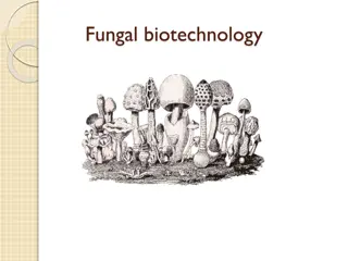
Characters of Fungi A Fungus (singular), Fungi (Plural). Fungi are eukaryotic plant-like organisms that lack chlorophyll. The study of fungi is called Mycology. Some fungi grow as a single-celled entity, termed yeast that grows either by a budding process or via binary fission. Threads of cells are called hyphae. Fungal hyphae repeatedly branch to form a network of filaments termed mycelium (sing., mycelia). Fungi are identified based on macro and microscopic characteristics. Molds are commonly cultured on fungal-selective or enriched media such as Saboraud Dextrose agar (SDA), Corn Meal agar, and Potato Dextrose agar (PDA).
Common spores of Asexual reproduction fungi The two most common types of asexual reproductive spores produced by fungi are conidiospores and sporangiospores. Conidiospores are borne externally in chains on an aerial hypha called a conidiophore.
Penicillium and Aspergillus are examples of molds that produce conidiospores. Penicilliumis one of the most common household molds and is a frequent food contaminant. The conidiosporesof Penicillium usually appear grey, green, or blue and are produced in chains on finger-like projections called phialidescoming off from the conidiophore. Aspergillus Aspergillus is another common contaminant. Although usually nonpathogenic, it may become opportunistic in the respiratory tract of a compromised host and, in certain foods, can produce mycotoxins. The conidiophore terminates in a ball-like structure called a vesicle. Its conidiospores, which typically appear brown to black, are produced in chains on phialides coming off from the vesicle.
Name of fungus Macro morphology Microscopic morphology (on agarplate) Penicillium
Name of fungus Macro morphology (on agarplate) Microscopic morphology Aspergillus
Sporangiospores are produced within a sac or sporangium on an aerial hypha called a sporangiophore. Rhizopusis an example of a mold that produces sporangiospores. Although usually nonpathogenic, it sometimes causes opportunistic o wounds and respiratory infections in the compromised host. At the end of its sporangiophore is dome-shaped end called a columella that extends into a sac-like structure called a sporangium. Its sporangiospores, typically brown or black, are produced within the sporangium . Anchoring structures called rhizoids are also produced on the vegetative hyphae.
Some common fungi Name of fungus Macro morphology Microscopic morphology (on agarplate) Rhizopus
Fusariumgrows fast with varying colony color depending on the isolates. Woolly to cottony, flat, spreading colonies. Within a few days, cover the entire agar plate. Conidia are the spores produced by Fusarium. There are two types- macro and micro-conidia, both of which can be seen under the microscope. Macroconidia are multi-celled, while microconidia are single - celled.
Name of fungus Macro morphology (on agar plate) Microscopic morphology Fusarium
Alternariadevelop and grow as elongated chains with conidiophores that are dark brown in color. In favorable conditions (moisture or rain, nutrition), spores, referred to as conidia, are produced (growing as buds from the conidiophores) from the conidiophores asexually.
Name of fungus Macro morphology (on agarplate) Microscopic morphology Alternaria
Dermatophytes The dermatophytes are a group of molds that cause superficial mycoses of the hair, skin, and nails and utilize the protein keratin, that is found in hair, skin, and nails, as a nitrogen and energy source. Infections are commonly referred to as ringworm or tinea infections. The three common Trichophyton, and Epidermophyton. These organisms grow well at 25 C. dermatophytes are Microsporum,
Home work 1- Answer in Word doc. 2- Give me a complete answer. 3- continue the previous report and fill the tables for results of the isolation of Microflora by describing the growth characters on both NA and PDA.