Respiratory Fungal Infections: Etiology, Diagnosis, and Treatment
Explore the complexities of respiratory fungal infections, including the etiology, common fungal species, risk factors, and classification of diseases such as aspergillosis. Learn about the challenges in diagnosing and treating these infections, which are less common but present significant difficulties compared to viral and bacterial infections.

Download Presentation

Please find below an Image/Link to download the presentation.
The content on the website is provided AS IS for your information and personal use only. It may not be sold, licensed, or shared on other websites without obtaining consent from the author. If you encounter any issues during the download, it is possible that the publisher has removed the file from their server.
You are allowed to download the files provided on this website for personal or commercial use, subject to the condition that they are used lawfully. All files are the property of their respective owners.
The content on the website is provided AS IS for your information and personal use only. It may not be sold, licensed, or shared on other websites without obtaining consent from the author.
E N D
Presentation Transcript
Lecture : Respiratory Fungal Infection important Extra notes Doctors notes " . "
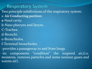
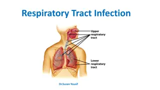
Objectives: Respiratory System Rout of infection? Oral Cavity, any role? Respiratory fungal infections are less common than viral and bacterial infections. Have significant difficulties in diagnosis and treatment.
Respiratory fungal infection - Etiology Dimorphic fungi (Primary infections) Common in travel YEAST Mould fungi (Opportunistic) (Opportunistic) Candidiasis (Candida) Aspergillosis (Aspergillus species) Histoplasma capsulatum Zygomycosis (y, e.g. Rhizopus, Mucor) Blastomyces dermatitidis Cryptococcosis Paracoccidioides brasiliensis (Cryptococcus neoformans, C. gattii) Other mould Coccidioides immitis
Primary Systemic Mycoses Infections of the respiratory system that occur by Inhalation. Dissemination seen in immunocompromised hosts. Common in North America and to a lesser extent in South America. Not common in other parts of the World. Etiologies are dimorphic fungi: In nature found in soil of restricted habitats. Primary pathogens, dimorphic fungi They are highly infectious They include: Histoplasmosis, Blastomycosis, Coccidioidomycosis, Paracoccidioidomycosis
Aspergillosis Aspergillosis is a spectrum of diseases of humans and animals caused by members of the genus Aspergillus of mould fungi (Opportunistic). These include : (1) Mycotoxicosis (such alpha toxin which produced by Aspergillus) (2) Allergy (3) Colonization (without invasion and extension ) in preformed cavities (4) Invasive disease of lungs (5) Systemic and disseminated disease. Common airborne Fungi Aetiological Agents: Aspergillus species, common species are: A. fumigatus, (Glabellar) A. flavus, (Regional) A. niger, A. terreus A. nidulans. Aspergillus niger Aspergillus fumigatus
CLASSIFICATION OF ASPERGILLOSIS: Risk factors : Bone marrow/ organ transplantation AIDS Diabetes others Cancer: Leukemia, lymphoma,.. Etc Airways/nasal exposure to airborne Aspergillus Invasive aspergillosis Aspergilloma of lung Maxillary (sinus) aspergilloma Chronic aspergillosis Allergic: Allergic bronchopulmonary (ABPA) Allergic Aspergillus sinusitis Persistence without disease - colonisation of the airways or nose/sinuses
Aspergillosis: Chronic Aspergillosis (Colonizing aspergillosis) (Aspergilloma OR Aspergillus fungus ball) (Fungus Ball is a mass or clumping of hyphae +mucous + fibrin + cellular debris) usually inside cavity. Signs include: Cough, hemoptysis, variable fever Radiology will show mass in the lung , radiolucent (air) crescent Invasive pulmonaryAspergillosis Signs: Cough , hemoptysis, fever, Leukocytosis Radiology will show lesions with halo sign Simple (single) aspergilloma Invasive pulmonary aspergillosis Note the Halo sign Note the Air crescent
Allergic bronchopulmonary (ABPA) Also: Symptoms of Asthma Bronchial obstruction Fever, malaise Eosinophilia Wheezing +/- Skin test reactivity to Aspergillus Serum antibodies to Aspergillus Serum IgE > 1000 ng/ml Pulmonary infiltrates
Fungal sinusitis Clinical : Nasal polyps and other symptoms of sinusitis In immunocompromised, Could disseminate to eye craneum (Rhinocerebral) The most common cause in KSA is Aspergillus flavus In addition to Aspergillus, there are other fungi that can cause fungal sinusitis Aspergillus sinusitis has the same spectrum of Aspergillus disease in the lung Diagnosis Clinical and Radiology Histology Culture Precipitating antibodies useful in diagnosis Measurementof IgE level, RAST test* Treatment : depends on the type and severity of the disease and the immunological status of the patient * A radioallergosorbent test is a blood test using radioimmunoassay test to detect specific IgE antibodies, to determine the substances a subject is allergic to.
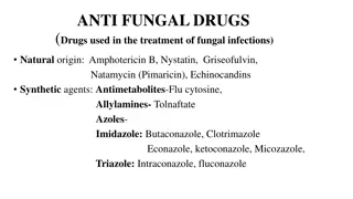
Diagnosis of Aspergillosis Specimen: Respiratory specimens: Sputum, BronchoAlveolar Lavage (BAL), Lung biopsy, Other samples: Blood, etc. Lab. Investigations: Direct Microscopy: Giemsa Stain, Grecott methenamine silver stain (GMS) Will show fungal septate hyphae Culture on SDA (Sabouraud Dextrose Agar is selective media) Serology: Test for Antibody ELISA test for galactomannan Antigen PCR: Detection of Aspergillus DNA in clinical samples Choice of antifungal for aspergillosis: Voriconazole Alternative therapy: Amphotericin B, Itraconazole, Caspofungin Usually we need surgery also
Zygomycosis: PulmonaryZygomycosis: o Pulmonary zygomycosis o Rhinocerebral zygomycosis o Acute infection o Consolidation , nodules, cavitation, pleural effusion, hemoptysis o Infection may extend to chest wall, diaphragm, pericardium. Pulmonary infractions and hemorrhage Rapid evolving clinical course o Early recognition and intervention are critical Risk factors Transplant patients Malignancy AIDS Diabetic ketoacidosis Many others Etiology: Zygomycetes , Non-septate hyphae e.g. Rhizopus, Note : Aspergillosis septate hyphae p.Zygomycosis non-septate hyphae
Diagnosis: Specimen: Respiratory specimens: Sputum, BAL, Lung biopsy, Other samples Lab. Investigations: Direct Microscopy: Giemsa, Grecott methenamine silver stain (GMS) Will show broad non- septate fungal hyphae Culture on SDA (no cycloheximide)because it may inhibit the growing of this fungi Serology: Not available Treatment: Amphotericin B Surgery
Pneumocystosis (PCP): Pneumocystis pneumonia (PCP) Opportunistic fungal pneumonia It is interstitial pneumonia of the alveolar area that effect immunocompromised hosts. Affect compromised host Especially common in AIDS patients. o Previously thought to be a protozoan parasite, but later it has been proven to be a fungus Etiology: Pneumocystis jiroveci Does not grow in laboratory media e.g. SDA ( only we can use silver \ IF stain which is more sensitive) Naturally found in rodents (rats), other animals (goats, horses), Humans may contract it during childhood Important slide
Pneumocystosis: Laboratory Diagnosis: Patient specimen: Bronchoscopic specimens (B.A.L.), Sputum (not very good), Lung biopsy tissue. Histological sections or smears stained by GMS stain. Immunuofluorescence (better sensitivity) If positive will see cysts of hat-shape, cup shape, crescent. Treatment: Trimethoprim sulfamethoxazole (the two drugs are combined and known as co-trimoxazole) Dapsone Important slide
Pulmonary Candidiasis Candida causing Pneumonia but Not Common. Has Primary and Secondary Secondary Dissimination of blood Diagnose: don t depend on Sputum Treatment: Fluconazole Voticonazole if resistant to fluconazole
summary: Opportunistic Primary infections Yeast Mould Fungi Dimorphic fungi Etiology Candidiasis (candida, and other yeast) Aspergillosis (Aspergillus species) Histoplasma capsulatum Respiratory Fungal Infections Blastomyces dermatitidis Zygomycosis (Zygomycetes eg. Rhizopus, mucor) Cryptococcosis (Cryptococcus neoformans, C. gatti) Paracoccidiodes brasiliensis Other mould Coccidioides immitis (1) Primary Systemic Mycoses (3) Zygomycosis (4) Pneumocystosis (PCP) Rhinocerebral zygomycosis Pulmonary zygomycosis (Acute) Etiology: Zygomycetes (rhizopus) Diagnosis: Specimen (sputum, BAL, biopsy) Lab (direct microscopy: GMS and Giemsa stain = broad non-septate fungal hyphae) (culture = SDA) (serology= not available) Treatment: Amphotericin B Surgery (Inhalation) Primary pathogens Highly infectious Etiology: Dimorphic fungi: Histoplasmosis Blastomycosis Coccidioidomycosis Paracoccidioidomycosis Opportunistic fungal pneumonia Etiology: Pneumocystis jiroveci Diagnosis: Specimen (sputum, BAL, biopsy) Histology (GMS stain) Immunofluorescence Treatment: Trimethoprim Sulfamethoxazole Dapsone Classification (2) Aspergillosis (next slide)
summary: (2) Aspergillosis Etiology: (Aspergillus species) A. fumigatus A. flavus A. niger A. terreus A. nidulans Diagnosis: Specimen (Sputum, BAL, biopsy, blood, etc) Lab (direct microscopy: GMS and Giemsa stain = septate hyphae) fungal (culture = SDA) (Serology = test for antibody, ELISA) (PCR = detection of aspergillus DNA in clinical sample) Treatment: Voriconazole Alternative: Amphotericin B, Itraconazole, Caspofungin Invasive aspergillosis Allergic Allergic aspergillus sinusitis Allergic bronchopulmonary Aspergillosis (ABPA): Symptoms of asthma, bronchial obstruction, fever, malaise, eosinophilia, wheezing +/- Signs: cough, hemoptysis, fever, leukocytosis. Radiology: Lesions with halo signs Chronic aspergillosis (colonizing apergillosis) Aspergilloma of lung Maxillary (sinus) aspergilloma Signs: cough, hemoptysis, variable fever Radiology: mass in the lung, radiolucent crescent Fungal Sinusitis Signs: nasal polyps and other symptoms of sinusitis Most common cause: Aspergillus flavus Diagnosis: clinical and radiology, histology, culture, RAST, IgE levels Treatment: Varies according to severity and type of the disease and the immunological status of the patient Persistence without disease colonization of the airways or nose/sinuses
GOOD LUCK! MICROBIOLOGY TEAM: Waleed Aljamal (leader) Ibraheem Aldeeri Ibrahim Fetyani Abdulaziz almohammed Abdulmalik alghannam Omar albabtain Turki maddi Mohammad alkahil Meshal Eiaidi Khalid Alhusainan Khalid Alshehri Nasir Aldosarie Shrooq Alsomali and Ghadah Almazrou (leaders) Reem Alshathri Ruba Barnawi Jawaher Abanumy Ouhod Abdullah Rawan alqahtani The Editing File We are waiting for your feedback @microbio436 436microbiologyteam@gmail.com


")


:")