Understanding Different Types of Anaemia in Veterinary Medicine
Explore the various types of anaemia in veterinary medicine, including haemorrhagic anaemia, haemolytic anaemia, nutritional anaemia, and anaemia due to immunological reactions. Learn about their causes, symptoms, and classifications to better diagnose and treat animals suffering from anaemia.

Download Presentation

Please find below an Image/Link to download the presentation.
The content on the website is provided AS IS for your information and personal use only. It may not be sold, licensed, or shared on other websites without obtaining consent from the author. If you encounter any issues during the download, it is possible that the publisher has removed the file from their server.
You are allowed to download the files provided on this website for personal or commercial use, subject to the condition that they are used lawfully. All files are the property of their respective owners.
The content on the website is provided AS IS for your information and personal use only. It may not be sold, licensed, or shared on other websites without obtaining consent from the author.
E N D
Presentation Transcript
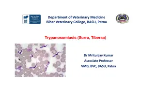
Department of Veterinary Medicine Bihar Veterinary College, BASU, Patna Anaemina Dr Mritunjay Kumar Associate Professor VMD, BVC, BASU, Patna
Anaemia is defined as reduction in the amount of haemoglobin per unit of blood and may or may not be accompanied with the reduction in red blood cells and characterised with paleness of mucous membranes, tachycardia and loss of muscular strength and vigour Aetiology Classification based on aetiology a) Haemorrhagic anaemia: Excessive blood loss and its severity depends on the rate of blood loss, amount of blood loss and rate of its production in body It can be either acute or chronic. i) Acute haemorrhagic anaemia: Severe wound injury, epistaxis, haemoptysis, surgical bleeding, splenomegaly in dogs, purpura haemorrhagica, braken fern poisoning, warfarin poisoning, rapid exposure to x-ray etc. ii) Chronic haemorrhagic anaemia: Ecto or endoparasitic infection causing sucking of blood, haemophilia, enzotic bovine haematuria, coccidial infection in young animals and deficiency of vitamin K and C and prothrombin
b) Haemolytic anaemia: Internal vascular haemolysis and normocytic macrocytic initially and become hypochromic microcytic when iron stores are utilised. Erythrocytes become more fragile and reveal spherocytosis. It may be either congenital or acquired. i) Congenital haemolytic anaemia: Dogs and cats as result of congenital defects in stroma production in protein or haemoglobin Bovine and swine congenital porphyria occurs. In it the level of porphyrin is photosensitive due to lack of enzyme that converts porphyrin into haemoglobin. Porphyrin is photosensitive and may cause pink tooth or osteohaemochromatosis when deposited in teeth or bone. i) Acquired haemolytic anaemia: Bacillary haemoglobinuria, leptospirosis, haemolytic streptococcal and staphylococcal infections, equine infectious anaemia, babesiosis, theileriosis, anaplasmosis, eperythrozoonosis, PPH and poisoning with copper, pheothiazine, arsenic, bismuth, lead or sulphonamides and snake venoms It is also recorded in haemonchosis, fascioliasis, coccidiosis In young calves or lambs prlonged use of oxytetracyclines and excessive consumption of Brassica species of plants.
c) Nutritional anaemia Deficiency of copper, cobalt, iron, niacin, riboflavin, pantothenic acid, pyridoxine and choline. d) Anaemia due to immunological reaction: i) Autoimmune haemolytic anemia: Erythrocytes are recognised as non-self and antibodies are produced against these cells results in antigen antibody reaction and haemolysis It is characterised by sudden onset of anaemia and spherocytosis and usually seen in dogs and horses i) Isoimmune haemolytic anaemia: It is usually seen in new borne animals and occurs due to transfer of maternal isoantibodies to them through colostrums. The disease can be seen in newborn calf whose dam has been repeatedly immunised against babesiosis, in foals following immunization of mare with equine viral abortion vaccine or in piglet after immunisation of swine with swine fever vaccine
e) Dyshaemopoetic anaemia: In chronic suppurative conditions bone marrow activity is suppressed leads to depression of erythropoiesis and less RBC production in nephritis, chronic interstitial nephritis, neoplastic diseases, aplasia of bone marrow, rapid exposure to x-rays or other radioactive substances, drug toxicity that suppress the bone marrow activity like sulphonamides, chloramphenicol and in bone marrow suppressing viral infection like parvo-virus infection Anaemia classification on morphological features a) Normocytic normochromic anaemia: It is also known as aplastic or hypoplastic anaemia as bone marrow activity is depressed. Here erythrocytes have normal staining properties but number of granulocytes and thrombocytes is reduced. It is primary or secondary but usually secondary anaemia is seen and primary is rarely recorded in domestic animals. Chronic haemorrhage, neoplasm, deficiency of vitamin B12 and prothrombin, irradiation, chemical poisoning, braken fern poisoning and excess use of sulphonamide and chloramphenicol b) Normocytic hypochromic anaemia: This type of anaemia is usually due to deficiency of some nutrients, deficiency of iron in diet or its defective absorption from intestine or deficiency of copper, ascorbic acid, pyridoxine, nicotinic acid, riboflavin or thyroxin in diet. c) Macrocytic normochromic anaemia: It occurs due to the presence immature erythrocytes in blood as a result of deficiency of vitamin B12 (RBC maturation), cobalt, folic acid (RBC maturation), intrinsic factors (needed for absorption of vitamin B12) and erythrocytes maturation factors
d) Macrocytic hypochromic anaemia: This type of anaemia occurs in regenerative phase after haemorrhage It may be noticed due to trauma, severe wound, surgical operations, epistaxis and infection with parasitic infections that result in oozing of blood e) Microcytic normochromic anaemia: It can be seen in chronic diseases like tuberculosis, brucellosis, chronic interstitial nephritis, parasitic diseases, metal poisoning, sulphonamides and chemical due to exposure to the irradiation f) Microcytic hypochromic anaemia It is also referred as iron deficiency anaemia and occurs as results of deficiency of copper, cobalt, manganese, ascorbic acid, pyridoxine, riboflavin and nicotinic acid
Pathogenesis Irrespective of the cause of anaemia there is development of anaemic anoxia The oxygen carrying capacity of blood is reduced due to low haemoglobin concentration Of the various tissues skeletal, cardiac and muscles and nervous tissue are affected maximum because oxygen demand of these tissues is more For maintaining oxygen supply speed of circulation to tissues is enhanced If such compensatory adjustments fail to maintain oxygen demand, cardiac failure may develop
In acute haemorrhagic anaemia there is loss of circulatory blood volume and plasma proteins If haemorrhage stops, the lost fluid is quickly restored by release of fluid from tissue spaces while plasma proteins are restored by their greater synthesis in liver In haemolytic anaemia, haemoglobinuria develops, followed by nephrosis and depressed renal function Aplastic anaemia occurs in toxicity and in reversed with the removal of cause Similarly nutritional anaemia is cured with the supplementation of desired nutrition in the diet
Clinical Findings In animals suffering from anaemia muscular weakness, dullness, depression and low working efficiency Such animal reveal inappetence or anorexia and get exhausted quickly The visible mucous membranes become pale, elevated heart rate and pulse as greater amplitude Intensity of heart sound is greatly increased due to increase in blood pressure and cardiac dilatation In later stages intensity of heart sound is decreased due to myocardial asthenia Due to cardiac dilation haemic systolic murmurs are audible They go in line with each respiratory cycle
In initial stage of disease signs of dyspnoea are not seen but at terminal stage dyspnoea occurs due to development of anaemic anoxia In haemorrhagic anaemia signs of shock are visible Jaundice, haemoglobinuria, oedema and haematuria may also noticed in some cases Necropsy findings In post-mortem examination carcass and most of the tissues appear pale in colour There is thin watery blood, and size of spleen is reduced In addition to these signs specific lesions of primary disease may also be seen. Diagnosis History and Clinical signs: History of specific etiological factors and presence of specific clinical Blood examination: Reduced Hb, TEC, PCV values, reduced total protein in haemorrhagic anaemia Calculations of MCV, MCH and MCHC helps to know the type of anaemia and involvement of the causal factor
MCV (Mean Corpuscular Volume) Definition: Average volume (size) of a single red blood cell Formula: MCV ( Fl) = (Hematocrit %)/(RBC x 10 12 /L) x 10 Normal Range (species-dependent): Dog: 60 77 fL Cat: 39 55 fL Clinical Significance: MCV (macrocytic): Regenerative anemia (e.g., blood loss, hemolysis), vitamin B12/folate deficiency MCV (microcytic): Iron deficiency anemia, chronic blood loss Normal MCV: Normocytic anemia (e.g., anemia of chronic disease)
MCH (Mean Corpuscular Hemoglobin) Definition: Average amount of hemoglobin in a single red blood cell Formula: MCH (pg) = (Hemoglobin in g/dL)/(RBC x 1012/L) x 10 Normal Range: Dog: 19 23 pg Cat: 13 17 pg Clinical Significance: MCH: Hypochromic anemia (iron deficiency) Normal MCH: Normochromic anemia MCH: Rare, may indicate lab artifact or macrocytic cells
MCHC (Mean Corpuscular Hemoglobin Concentration) Definition: Average concentration of hemoglobin in a given volume of packed red blood cells. Formula: MCHC (g/dl) = (Hemoglobin in g/dL)/ (Hematocrit %) x 100 Normal Range: Dog: 32 36 g/dL Cat: 30 36 g/dL Clinical Significance: MCHC (hypochromic): Iron deficiency anemia MCHC: Usually artifactual (e.g., hemolysis, lipemia)
Summary Table: Parameter Definition Formula Significance Microcytic, normocytic, macrocytic MCV ( Fl) Size of RBC (PCV 10) / RBC Hypochromic, normochromic MCH (pg) Hemoglobin per RBC (Hb 10) / RBC Hypochromic, (pseudo)hyperchromic MCHC (g/dl) Hb concentration in RBCs (Hb 100) / PCV
Treatment Treat the primary cause of disease Blood transfusion in acute haemorrhage and chronic anaemia of severe degree The plasma expanders like haemocoel, Vetplasma, dextran or gelatine polymers may also be used as substitute of whole blood. Along with these therapy hematinic preparations like iron dextran may be given orally Mineral and vitamin supplementation of the diet helps in recovery of cases suffering from anaemia of nutritional origin For management of cases of autoimmune haemolytic anaemia corticosteroids should be given
Erythropoiesis-Stimulating Agents (ESAs) These agents stimulate red blood cell production and are primarily used for anemia associated with chronic kidney disease or chemotherapy 1. Epoetin Alfa Mechanism: Recombinant human erythropoietin that mimics endogenous erythropoietin Dosage: Dogs and cats: Initial dose of 50-100 U/kg, subcutaneously (SC), three times per week until the low end of the target packed cell volume (PCV) range is reached; then, frequency decreased to once weekly. Trade Names: Epogen, Abseamed, Retacrit, Binocrit, Zyrop Adverse Effects: Autoantibody development leading to treatment resistance and worsening anemia (pure red cell aplasia) is the most serious adverse effect 2. Darbepoetin Alfa Mechanism: Synthetic form of erythropoietin with a threefold longer half-life Dosage: Dogs: Initial dose of 0.45 1.5 mcg/kg, SC, once weekly until the low end of the target PCV range is reached; then, dosing interval extended as tolerated. Cats: Initial dose of 0.7 1.8 mcg/kg, SC, once weekly until the low end of the target PCV range is reached; then, dosing interval extended as tolerated. Trade Names: Aranesp (Amgen), Cresp (Dr. Reddy s Laboratories in India).
Hematinics (Supportive Therapy) These agents support red blood cell production and are used for various types of anemia, including those due to nutritional deficiencies. 1. Vitamin B (Cobalamin, Cyanocobalamin) Role: Essential for DNA synthesis and red blood cell maturation Indications: Macrocytic anemia due to deficiencies Dosage: Parenteral: 20 50 mcg/kg, SC, once weekly for 4 6 weeks; then monthly. Oral: 50 mcg/kg, orally, once daily for at least 12 weeks. 2. Folic Acid (Vitamin B ) Role: Essential for DNA and RNA synthesis. Indications: Macrocytic anemia due to deficiencies. Dosage: Dogs: 5 mg, orally, once daily for 1 month. Cats: 2.5 mg, orally, once daily for 1 month. Injectable: 1 5 mg, SC, once weekly for 1 month.
Iron Role: Necessary for hemoglobin formation Indications: Microcytic, hypochromic anemia due to iron deficiency Dosage: Parenteral: Dogs: 10 20 mg/kg, intramuscularly (IM), once; maximum 300 mg/dog Cats: 10 mg/kg, IM, once Oral Dogs: 100 300 mg/day Cats: 50 100 mg/day


 Haemolytic anaemia: Internal vascular haemolysis and")
 Nutritional anaemia")
 Dyshaemopoetic anaemia: In chronic suppurative conditions")
 Macrocytic hypochromic anaemia:")


")
")
")


")
")


![Enhancing Patient Blood Management in Pregnancy at [INSERT LOCAL HOSPITAL NAME]](/thumb/143207/enhancing-patient-blood-management-in-pregnancy-at-insert-local-hospital-name.jpg)