Anaemia: Symptoms, Diagnosis, and Treatment
Explore the comprehensive guide on anaemia, a condition affecting billions worldwide. Learn about the causes, symptoms, and diagnostic approaches for different types of anaemia, such as iron deficiency anaemia and microcytic anaemia. Find insights on the importance of history, examination, and laboratory tests in identifying and managing anaemia effectively.

Download Presentation

Please find below an Image/Link to download the presentation.
The content on the website is provided AS IS for your information and personal use only. It may not be sold, licensed, or shared on other websites without obtaining consent from the author. If you encounter any issues during the download, it is possible that the publisher has removed the file from their server.
You are allowed to download the files provided on this website for personal or commercial use, subject to the condition that they are used lawfully. All files are the property of their respective owners.
The content on the website is provided AS IS for your information and personal use only. It may not be sold, licensed, or shared on other websites without obtaining consent from the author.
E N D
Presentation Transcript
ANAEMIA Dr C du Toit 2020
Anaemia 2 Billion people in world are anaemic Hemoglobin below the normal range for age gender (and altitude) Not a diagnosis a clinical / laboratory finding of which the cause must be found History Examination Laboratory
Anaemia History -symptoms related to the anaemia -severity -time period over which drop in hemoglobin occurred chronic vs acute B12 deficiency vs rupture of ectopic pregnancy -symptoms related to the cause of the anaemia Examination -features of the anaemia conjunctival pallor, ejection systolic murmur -features of the cause of the anaemia epigastric tenderness and melena stools on glove
Anaemia FBC Low haemoglobin confirms clinical suspicion Reticulocyte count high = increased red cell loss or destruction -bleeding -hemolysis low = decreased production -iron /folate deficiency, chronic disease
Anaemia MCV Microcytic Normocytic Macrocytic
Case 1 A 22 year old woman presents with shortness of breath and palpitations. She has a history of heavy irregular menstrual bleeding. She is anaemic. The Hb =7g/dl with platelets 650x10 9/l ,a normal white cell count. MCV = 68 fl.
Microcytic anaemia Differential diagnosis Iron deficiency Anaemia of chronic disease Thalassemia Lead poisoning Sideroblastic anaemia
Iron deficiency One of the most common medical conditions Any age or gender Symptoms of chronic anaemia , may complain of blood loss or possible causes Examination may indicate cause Fbc Low Hb, Low MCV white cells normal Platelets high or normal rarely low Smear
Iron deficiency. Note the red cells are smaller than the nucleus of the lymphocyte indicated by the arrow. The red cells have a large area of central pallor with a rim of Haemoglobin. The arrow on the right indicates a pencil cell- typical of iron deficiency
Iron deficiency Iron studies to confirm suspicion s= ferritin low = iron deficiency can be false normal with infection/ inflammation transferrin levels High in iron deficiency transferrin saturation Low in iron deficiency
Iron deficiency Management - 2 steps Find and treat cause of iron loss /deficiency Gynaecology evaluation, GIT scopes , rarely form urinary tract Supplement the iron -oral ferrosulphate preferred GIT side effects common can improve absorption by dosing alternate days or even just once a day Other oral preparations, less side effects ,more cost -Iv iron Venofer / Cosmofer Cost Risk of allergic reactions Iv iron used more often give pre surgery rather than transfusions and for patients who struggle to tolerate iron or where the iron loss cannot be stopped
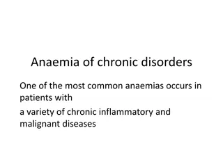
Anaemia of chronic disease Mild to moderate anaemia seldom <7g/dl Starts normocytic becomes microcytic if long duration Main mechanisms of the anaemia: inflammatory cytokines Increased hepcidin release by liver Internalisation of ferroportin in cells Inability to release iron to the bone marrow Shortened red cell survival and decreased sensitivity of marrow to erythropoietin also plays a part.
Thalassemia Group of inherited conditions with abnormalities of Hemoglobin Rare in South Africa Very low MVC with target cells on smear Most common types are: thalassemia with decrease or absence of chains and excess chains - thalassemia with varying levels of chains Carriers (thalassemia minor) may have mild anaemia or normal Hb Thalassemia major transfusion dependent from 6 months old risk of iron overload needing life-long chelation.
Case 2 A 65 year old woman who is being investigated for early dementia is found to be anaemic. The FBC shows : Hb =8g/dl, white cells =2.5x 10 9 /l and platelets =110x10 9/l. The MCV is 116 fl.
Macrocytic anaemia MCV >100 2 Main groups Round macrocytes Liver disease Hypothyroidism Pregnancy High reticulocyte count (bigger than normal red cells) Megaloblastic anaemia (Oval macrocytes) (ineffective DNA production and cell division) B12 deficiency Folate deficiency
Megaloblastic anaemia Clinically pale with mild jaundice B12 deficiency patients may have peripheral neuropathy or in severe cases spastic paraparesis and dementia Full blood count may range form megaloblastic anaemia only to pancytopenia due to ineffective DNA production Smear shows oval macrocytes and neutrophils with hypersegmented nuclei. Chemistry very high LDH(>1000 often 4000+) due to ineffective erythropoiesis in marrow with cell lysis
Megaloblastic anaemia. Note the large oval shaped red cells and the hyper segmented neutrophil nuclei
B12 deficiency Remember the risk of irreversible paraparesis and dementia if not treated Stores last for up to 3 years -deficiency mostly due to malabsorption pernicious anaemia total gastrectomy pathology of terminal ileum non specific malabsorption in elderly (medication) -only strict vegans deficient due to diet Patients with malabsorption will need life-long monthly B12 injections
Folate deficiency Stores last for 4 months only Deficiency often due to -low intake (rare with food fortification) -increased requirements (pregnancy, haemolysis) -drugs -rarely malabsorption
Acute Management Draw blood for S B12 and s-folate Start treatment with B12 injections and oral folic acid Never give folate alone may improve anaemia even in B12 deficiency can lead to irreversible neurological damage Do not transfuse unless in failure one unit with diuretic cover Monitor for hypokalaemia
Haemolytic anaemia Decreased red cell survival due to destruction BM can compensate up to red cell survival of 15- 20 days Diagnosing haemolytic anaemia always follow the steps: Suspect haemolysis sudden drop in HB not due to bleeding anaemia and jaundice or splenomegaly family history of anaemia anaemia in Lupus/ other conditions associated with haemolysis
Haemolytic anaemia Confirm haemolysis High LDH and haptoglobin High reticulocyte count Find the cause start with: Smear Coombs
Spherocytes Blister cells and bite cells G6PD deficiency Sickle cells
Haemolysis causes Red cell abnormalities (Inherited) Causes outside the red cell (Acquired) -Autoimmune Warm antibody Cold antibody Drugs -Fragmentation Valves MAHA -Membrane Spherocytosis -Enzyme G6pd Deficiency -Hemoglobin Sickle cell disease -Hypersplenism -Infections Malaria Clostridia -Toxins -PNH
Case3 A 35 year old man with HIV who recently started ante- retroviral drugs presents with severe tiredness and shortness of breath. On examination he has severe anaemia with no other significant findings. The Hb =5g/dl, MCV 85 fl with normal white cells and platelets. The reticulocyte count is 0. The bone marrow shows a block in red cell production a early level. Pure red cell aplasia Causes in HIV -Parvovirus do PCR -ARV S lamivudine, rarely emtricitabine
Pure red cell aplasia due to ParvoB19 giant pronormoblast


![Enhancing Patient Blood Management in Pregnancy at [INSERT LOCAL HOSPITAL NAME]](/thumb/143207/enhancing-patient-blood-management-in-pregnancy-at-insert-local-hospital-name.jpg)