Learn about ulcerative lymphangitis in horses, a bacterial infection affecting lymphatic vessels. Discover its clinical signs, manifestations, and diagnosis methods, along with transmission and host range information.
Please find below an Image/Link to download the presentation.
The content on the website is provided AS IS for your information and personal use only. It may not be sold, licensed, or shared on other websites without obtaining consent from the author. If you encounter any issues during the download, it is possible that the publisher has removed the file from their server.
You are allowed to download the files provided on this website for personal or commercial use, subject to the condition that they are used lawfully. All files are the property of their respective owners.
The content on the website is provided AS IS for your information and personal use only. It may not be sold, licensed, or shared on other websites without obtaining consent from the author.
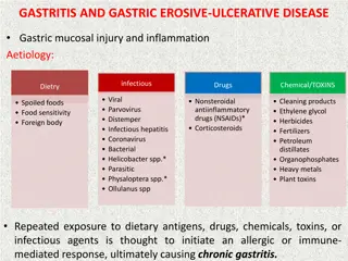
ULCERATIVE LYMPHANGITIS Synonym: Pigeon Fever in Horse Ulcerative lymphangitis is a sequel to bacterial infection(s) of the cutaneous lymphatic vessels of Equines, ultimately affects the performance of horse due to lameness and deformity of the limbs. The organism produces various extracellular exotoxins, which play a role in virulence. Etiology:Corynebacterium pseudotuberculosis Host Range: Equids, sheep, goats, cattle, buffalo, camelids, and rarely humans. Clinical Signs: Three forms have been described in horses: Limb infection/Ulcerative lymphangitis External abscesses Enternal infection
Limb infection: i. There is severe limb swelling and cellulitis, with multiple draining tracts lymphatics. ii. One or both hind limbs are affected cause severe lameness, fever, lethargy and anorexia. iii. In chronic stage , resulting in limb oedema, prolonged or recurrent infection, lameness, weakness, and weight loss following Ulcerative lymphangitis
External Abscesses: i. Most common manifestation ii. a single or multiple abscesses can occur anywhere on the body (prepuce, mammary gland, triceps, axilla, limbs, and head), frequently develop in the pectoral region along the ventral midline abdomen and appears as a pigeon s breast. iii. Abscess may ooze odor-free purulent exudate and are usually well encapsulated. iv. External abscesses do not usually develop signs of systemic illness but most of swelling the Fig. : The bursting nodules can discharge pus
Internal infection: i. Occurs in approximately 8% of affected horses ii. The organs most commonly involved are liver, kidney, spleen and lungs. iii. Pregnant foal may abort due to placentitis or fetal infection The most common clinical signs to be remembered concurrent external abscesses decreased appetite fever, lethargy weight loss Signs of respiratory disease or abdominal pain. Other signs observed in horses with internal abscesses include ventral oedema, ventral dermatitis, ataxia, hematuria (due to renal abscesses), and uncommonly, abortion.
Transmission: i. It may be transmitted through horse-to-horse contact or from infected to susceptible horses via insects, other vectors, or contaminated soil ii. The portal of entry is through abrasions or wounds in the skin or mucous membranes. iii. Mechanical vectors (Haematobia irritans, Musca domestica, Stomoxys calcitrans) iv. ventral midline dermatitis is a predisposing cause of infection. Diagnosis: i. Bacterial culture of aspirates or exudate ii. Clinical pathological abnormalities: anemia of chronic disease, leukocytosis with neutrophilia, hyperfibrinogenemia, and hyperproteinemia.
iii. Diagnosis of internal infection is based on clinical signs, clinicopathological data, serology, diagnostic imaging and bacterial culture. iv. Serologic testing: Synergistic Hemolysis Inhibition (SHI), may aid in diagnosis of internal abscesses. Treatment: i. Drainage of external abscesses ii. Antimicrobial therapy as penicillin G, macrolides, tetracyclines, cephalosporins, fluoroquinolones and rifampin may be recommondded. Trimethoprim-sulfa (5 mg/kg based on the trimethoprim fraction, twice daily orally) or chloramphenicol
Procaine intramuscularly) are effective for external abscesses especially on the ventral midline. Rifampin (2.5-5 mg/kg twice daily orally) in combination with ceftiofur (2.5 5 mg/kg twice daily intramuscularly) appears highly effective for treatment of internal abscesses. Internal abscesses have also respond well to enrofloxacin (7.5 mg/kg once daily orally). The average duration of antimicrobial therapy for internal infection is 4-6 weeks which is best determined by repeat abdominal ultrasound and clinic pathologic results. Physical therapy (hydrotherapy, hand walking, and leg wraps)as well as NSAIDs are also recommended. penicillin (20,000 U/kg twice daily
Zoonotic Potential: Human illness through working with infected sheep developed axillary lymphadenitis. Prevention and Control: Wearing of gloves when working with affected horses followed by hand washing is indicated. Isolation of affected horses from naive herd mates Protecting horses from insect exposure by regular application of insect repellents. Meticulous wound care (topical fly repellents, antimicrobial ointments and bandages) to prevent infection from a contaminated environment